Review
. 2021 Nov 15;9(1):6-19.
doi: 10.1002/mdc3.13354. eCollection 2022 Jan.

Pragmatic Approach on Neuroimaging Techniques for the Differential Diagnosis of Parkinsonisms

Affiliations
Review

Pragmatic Approach on Neuroimaging Techniques for the Differential Diagnosis of Parkinsonisms

Cecilia Peralta et al. Mov Disord Clin Pract. .

Abstract

Background: Rapid advances in neuroimaging technologies in the exploration of the living human brain also apply to movement disorders. However, the accurate diagnosis of Parkinson's disease (PD) and atypical parkinsonian disorders (APDs) still remains a challenge in daily practice.

Methods: We review the literature and our own experience as the Movement Disorder Society-Neuroimaging Study Group in Movement Disorders with the aim of providing a practical approach to the use of imaging technologies in the clinical setting.

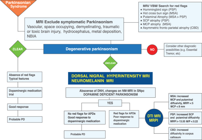
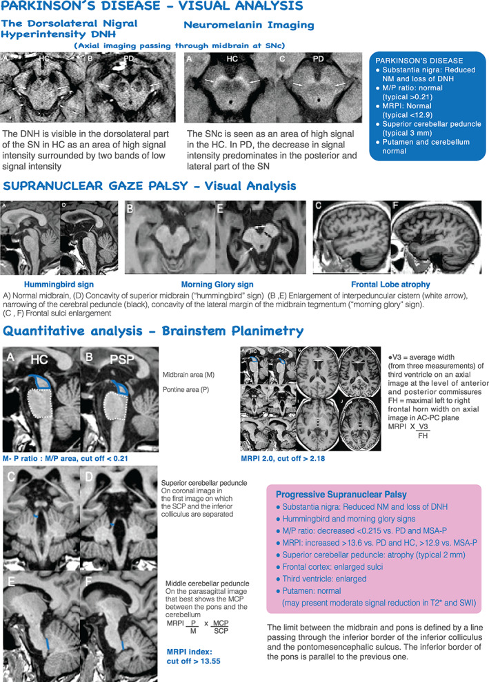
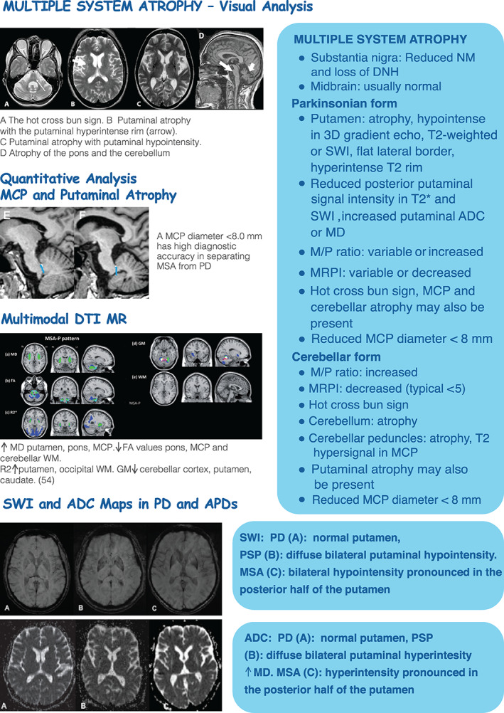
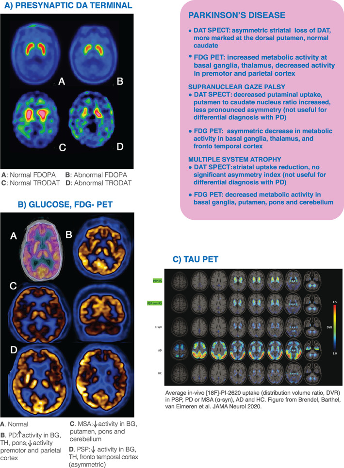
Results: The enormous amount of articles published so far and our increasing recognition of imaging technologies contrast with a lack of imaging protocols and updated algorithms for differential diagnosis. The distinctive pathological involvement in different brain structures and the correlation with imaging findings obtained with magnetic resonance, positron emission tomography, or single-photon emission computed tomography illustrate what qualitative and quantitative measures may be useful in the clinical setting.

Conclusion: We delineate a pragmatic approach to discuss imaging technologies, updated imaging algorithms, and their implications for differential diagnoses in PD and APDs.

Keywords: MRI; PET; Parkinson's disease; atypical parkinsonisms; imaging; multiple system atrophy; progressive supranuclear palsy.


Conflict of interest statement

Antonio P. Strafella is supported by the Canada Research Chair program and Canadian Institutes of Health Research (PJ8–1699695). Stephane Lehericy is supported by the Investissements d'Avenir (IAIHU‐06 Paris Institute of Neurosciences–Instituts hospitalo‐universitaires‐IHU and ANR‐11‐INBS‐0006) and Biogen Inc.

Figures

FIG 1
FIG 1
MR imaging diagnosis algorithm. APDs, atypical parkinsonian disorders; CBD, corticobasal degeneration; DNH, dorsal nigral hyperintensity; MCP, middle cerebellar peduncle; MR, magnetic resonance; MRI, magnetic resonance imaging; MRPI, Magnetic Resonance Parkinsonism Index; MSA, multiple system atrophy; NM, neuromelanin; PD, Parkinson's disease; PSP, progressive supranuclear palsy; SCP, superior cerebellar peduncle; NBIA, Neurodegeneration with Brain Iron Accumulation.
FIG 2
FIG 2
MR imaging in PD and PSP. DNH, dorsal nigral hyperintensity; HC, healthy control; MCP, middle cerebellar peduncle; MR, magnetic resonance; MRI, magnetic resonance imaging; MRPI, Magnetic Resonance Parkinsonism Index; NM, neuromelanin; PD, Parkinson's disease; PSP, progressive supranuclear palsy; SN, substantia nigra; SNc, substantia nigra pars compacta; SCP, superior cerebellar peduncle; SWI, susceptibility weighted imaging; AC‐PC, anterior commissure ‐ posterior commissure; MSA‐P, Multiple system atrophy‐ parkinsonian type; M/P, midbrain‐to‐pons ratio.
FIG 3
FIG 3
MR imaging in MSA and APDs. ADC, apparent diffusion coefficient; APDs, atypical parkinsonian disorders; DNH, dorsal nigral hyperintensity; FA, fractional anisotropy; HC, healthy control; MCP, middle cerebellar peduncle; MD, mean diffusivity; MR, magnetic resonance; MRI, magnetic resonance imaging; MRPI, Magnetic Resonance Parkinsonism Index; MSA, multiple system atrophy; NM, neuromelanin; PD, Parkinson's disease; PSP, progressive supranuclear palsy; SWI, susceptibility weighted imaging; VM, white matter; GM, gray matter; M/P, midbrain‐to‐pons ratio.
FIG 4
FIG 4
PET imaging of DA, Glucose and Tau protein aggregation. AD, Alzheimer's disease; APDs, atypical parkinsonian disorders; BG, basal ganglia; DAT‐SPECT, dopamine transporter single‐photon emission computed tomography; FDOPA, [18F]fluoro‐L‐dopa uptake; HC, healthy control; MR, magnetic resonance; MSA, multiple system atrophy; PD, Parkinson's disease; PET, positron emission tomography; PSP, progressive supranuclear palsy; TH, thalamus; FDG:18 Fluorodeoxyglucose; DA, dopaminergic; RS, Richardson´s Syndrome; TRODAT, technetium‐labeled dopamine transporter ligand. Fig 4C is reprinted from Brendel et al.
FIG 5
FIG 5
Multimodal imaging SWI MRI 3T‐ DAT SPECT. DAT‐SPECT, dopamine transporter single‐photon emission computed tomography; DNH, dorsal nigral hyperintensity; PET, positron emission tomography; SCP, superior cerebellar peduncle; SN, substantia nigra; SWI, susceptibility weighted imaging.
FIG 6
FIG 6
PET imaging diagnosis algorithm. APDs, atypical parkinsonian disorders; CBD, corticobasal degeneration; MSA, multiple system atrophy; PD, Parkinson's disease; PET, positron emission tomography; PSP, progressive supranuclear palsy; SPECT, single‐photon emission computed tomography; NBIA: Neurodegeneration with Brain Iron Accumulation.

Similar articles

Cited by

References

    1. Tondo G, Esposito M, Dervenoulas G, Wilson H, Politis M, Pagano G. Hybrid PET‐MRI applications in movement disorders. Int Rev Neurobiol 2019;144:211–257. - PubMed
    1. Rizzo G, Copetti M, Arcuti S, Martino D, Fontana A, Logroscino G. Accuracy of clinical diagnosis of Parkinson disease: a systematic review and meta‐analysis. Neurology 2016;86(6):566–576. - PubMed
    1. Adler CH, Beach T, Hentz J, et al. Low clinical diagnostic accuracy of early vs advanced Parkinson disease: a clinicopathologic study. Neurology 2014;83(5):406–412. - PMC - PubMed
    1. Fiford CM, Ridgway GR, Cash DM, et al. Patterns of progressive atrophy vary with age in Alzheimer's disease patients. Neurobiol Aging 2018;63:22–32. - PMC - PubMed
    1. Postuma RB, Berg D, Stern M, et al. MDS clinical diagnostic criteria for Parkinson's disease. Mov Disord 2015;30(12):1591–1560. - PubMed

LinkOut - more resources