. 2021 Dec 5;11(23):e4253.
doi: 10.21769/BioProtoc.4253.

A Phenotypic Screen for the Liver Stages of Plasmodium vivax

Affiliations

A Phenotypic Screen for the Liver Stages of Plasmodium vivax

Steven P Maher et al. Bio Protoc. .

Abstract

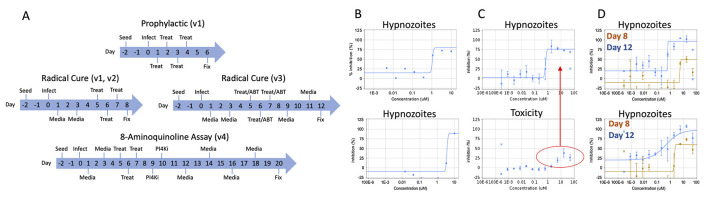
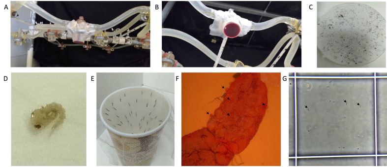
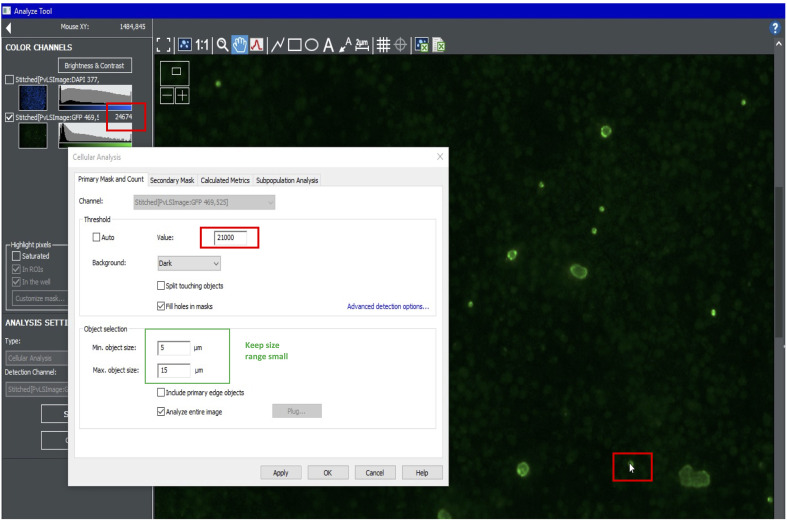
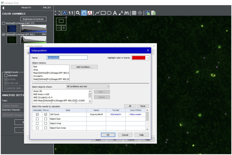
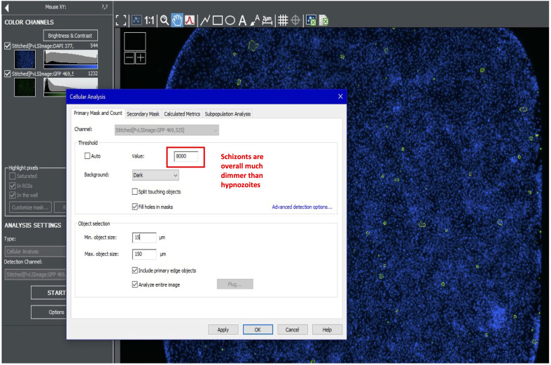
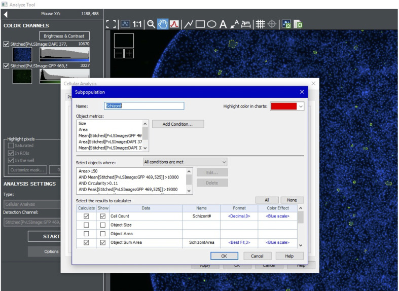
Control of malaria caused by Plasmodium vivax can be improved by the discovery and development of novel drugs against the parasite's liver stage, which includes relapse-causing hypnozoites. Several recent reports describe breakthroughs in the culture of the P. vivax liver stage in 384-well microtiter plates, with the goal of enabling a hypnozoite-focused drug screen. Herein we describe assay details, protocol developments, and different assay formats to interrogate the chemical sensitivity of the P. vivax liver stage in one such medium-throughput platform. The general assay protocol includes seeding of primary human hepatocytes which are infected with P. vivax sporozoites generated from the feeding of Anopheles dirus mosquitoes on patient isolate bloodmeals. This protocol is unique in that, after source drug plates are supplied, all culture-work steps have been optimized to preclude the need for automated liquid handling, thereby allowing the assay to be performed within resource-limited laboratories in malaria-endemic countries. Throughput is enhanced as complex culture methods, such as extracellular matrix overlays, multiple cell types in co-culture, or hepatic spheroids, are excluded as the workflow consists entirely of routine culture methods for adherent cells. Furthermore, installation of a high-content imager at the study site enables assay data to be read and transmitted with minimal logistical delays. Herein we detail distinct assay improvements which increase data quality, provide a means to limit the confounding effect of hepatic metabolism on assay data, and detect activity of compounds with a slow-clearance phenotype. Graphical abstract: Overview of P. vivax liver stage screening assay performed at the Institute Pasteur of Cambodia.

Keywords: 8-aminoquinolines; Antirelapse; Hypnozoites; Liver stage assay; Phenotypic screening; Plasmodium vivax; Primaquine; Tafenoquine.


Conflict of interest statement

Competing interestsThe authors have no competing interests to declare.

Figures

Figure 1.
Figure 1.. Overview of assay modes, versions, and example data.
A. In prophylactic assays, cultures are treated shortly after sporozoite infection of hepatocytes, in order to assess activity against immature hypnozoites and early schizonts. In radical cure assays, cultures are treated at 5 days post-sporozoite infection to allow for hypnozoites to mature. The v1 and v2 radical cure assays share the same timecourse, but feature different plate maps and normalization protocols. The v3 radical cure assay includes an extended endpoint (12 days post-infection) and addition of 1-aminobenzotriazole (ABT) to the medium on treatment days. The v4 assay includes a further extended endpoint of 20 days post-infection and addition of a PI4K (phosphatidylinositol 4-kinase) inhibitor (“PI4Ki”) to eliminate schizonts present at 9-10 days post-infection. “Seed” indicates hepatocyte seed, “Infect” indicates infection with sporozoites, “Media” indicates media change, “Treat” indicates compound treatment via the pin tool. The endpoint of all assays is fixation (“Fix”) prior to immunofluorescence staining and high content imaging. B. Example data from a v1 radical cure assay for two hit compounds with activity against hypnozoites. C. Example data from a v2 radical cure assay for a compound with nonselective activity against hypnozoites. The highest three doses of compound show a decrease in hepatic nuclei, indicating that cytotoxic conditions (red circle), not hypnozonticidal activity, are killing hypnozoites (top, red arrow) at those concentrations. Bars represent standard deviation (SD). D. Example data from v2 (orange) and v3 (blue) radical cure assays for two developmental compounds with activity against hypnozoites. The v2 curves show partial top plateaus that become complete inhibited with the longer v3 assay. Bars represent SD. Images and data are adapted from Maher et al. (2021) , used with permission. Figure 1A was adapted from Maher et al. (2021) and reproduced under a Creative Commons 4.0 license (http://creativecommons.org/licenses/by/4.0/).
Figure 2.
Figure 2.. Mosquito rearing and infection.
A. Side view of the apparatus for membrane feeding using a water-jacketed mini-feeder. B. Bottom view of a water-jacketed mini-feeder with a parafilm membrane holding blood. C. Whatman paper with mosquito eggs (a female An. dirus mosquito is included for size context). D. A single An. dirus pupae. E. A paper cup with a mesh lid used to contain females at day 5 post-emergence. F. Mosquito midgut stained with 1% mercurochrome in PBS following dissection at 7-days post-infectious bloodmeal, imaged under magnification with a 10× objective. Arrows show oocysts. G. A hemocytometer with P. vivax sporozoites harvested from An. dirus salivary glands at day 16 post-infectious bloodmeal, imaged under magnification with a 20× objective. The white box is 0.1 mm × 0.1 mm and arrows indicate three example sporozoites.
Video 1.
Video 1.. Occular view of a An. dirus midgut dissection to count oocysts.
Video 2.
Video 2.. Occular view of a An. dirus salivary gland dissection to obtain sporozoites.
Video 3.
Video 3.. Making a dilution series in a 384-well microtiter plate with a Biomek 4000.
Figure 3.
Figure 3.. Drug plate preparation, assay plate incubation, and spin holders.
A. An example drug plate following serial dilution using a Biomek 4000. B. Assay plates are stored in assay pans with four small Petri dishes holding water to maintain humidity and prevent edge effects during incubation in a cell culture incubator. The assay pans also help prevent introduction of contaminating microbes when plates are moved between incubators, microscopes, and biosafety cabinets. The white arrow indicates water in a Petri dish, and the yellow arrow indicates an assay plate. C. Design schematics for a custom-fabricated aluminum spin holder for removing spent media, values are mm. The design has been uploaded to the NIH 3D Print Exchange (https://3dprint.nih.gov/discover/3dpx-015808).
Video 4.
Video 4.. Mosquito wash and placement on slide prior to salivary gland dissection.
Figure 4.
Figure 4.. Overview of salivary gland dissection.
A. Mosquitoes designated for dissection are transferred to plastic cups covered in mesh and moved into the dissection room. B. Mosquito wash buffers are prepared in a 6-well plate on ice. C. Mosquitoes are prepared for dissection by first being sprayed with 70% ethanol and then transferred to the first well of a 6-well plate containing antibiotics (penicillin, streptomycin, and neomycin) in PBS. Individual mosquitoes are then washed in 70% ethanol and rinsed in PBS. E. Washed mosquitoes are placed facing the same direction on a sterile glass slide. Video 4 demonstrates how mosquitoes are washed. F. Slides are viewed under a stereomicroscope. Glands are dissected into an Eppendorf tube with RPMI on ice (blue box at top left). G. A dissector using a dissection needle (right hand) and forceps (left hand). Video 2 demonstrates how salivary glands are dissected.
Video 5.
Video 5.. Method for washing the Pin tool and transferring compounds into assay plates.
Figure 5.
Figure 5.. Pin tool wash and compound transfer.
The morning wash steps are used to prepare the pin tool for a treatment. After a treatment, the pin tool is washed and cleaned. After the last plate is treated on a given day, the tool is washed and cleaned again before storage overnight.
Figure 6.
Figure 6.. Setting the primary mask for hepatic nuclei.
Figure 7.
Figure 7.. Setting subpopulation parameters for hepatic nuclei.
Figure 8.
Figure 8.. Setting primary mask for hypnozoites.
Figure 9.
Figure 9.. Setting subpopulation parameters for hypnozoites.
Figure 10.
Figure 10.. Setting primary mask for schizonts.
Figure 11.
Figure 11.. Setting subpopulation parameters for schizonts.
Figure 12.
Figure 12.. Liver stage parasite size histograms and form classification.
The red box indicates the zoomed area of the top charts. Red and blue dots represent parasites from two different independent experiments. Hypnozoites are 28-100 µm2, but a small number of very large hypnozoites (or, possibly reactivating schizonts) are found between 100-150 µm2.
Figure 13.
Figure 13.. Example dose response curves and toxic point marking.
Figure 14.
Figure 14.. Demonstrated variability in sporozoite production, inoculum-based infection rate, and well-well infection rate.

References

    1. American Committee Of Medical Entomology American Society Of Tropical Medicine And Hygiene(2019). Arthropod Containment Guidelines, Version 3.2. Vector Borne Zoonotic Dis 19(3): 152-173. - PMC - PubMed
    1. Bousema T., Dinglasan R. R., Morlais I., Gouagna L. C., van Warmerdam T., Awono-Ambene P. H., Bonnet S., Diallo M., Coulibaly M., Tchuinkam T., et al. .(2012). Mosquito feeding assays to determine the infectiousness of naturally infected Plasmodium falciparum gametocyte carriers . PLoS One 7(8): e42821. - PMC - PubMed
    1. Chu C. S., Bancone G., Moore K. A., Win H. H., Thitipanawan N., Po C., Chowwiwat N., Raksapraidee R., Wilairisak P., Phyo A. P., et al. .(2017). Haemolysis in G6PD Heterozygous Females Treated with Primaquine for Plasmodium vivax Malaria: A Nested Cohort in a Trial of Radical Curative Regimens . PLoS Med 14(2): e1002224. - PMC - PubMed
    1. Chua A. C. Y., Ananthanarayanan A., Ong J. J. Y., Wong J. Y., Yip A., Singh N. H., Qu Y., Dembele L., McMillian M., Ubalee R., et al. .(2019). Hepatic spheroids used as an in vitro model to study malaria relapse . Biomaterials 216: 119221. - PubMed
    1. Dembele L., Franetich J. F., Lorthiois A., Gego A., Zeeman A. M., Kocken C. H., Le Grand R., Dereuddre-Bosquet N., van Gemert G. J., Sauerwein R., et al. .(2014). Persistence and activation of malaria hypnozoites in long-term primary hepatocyte cultures. Nat Med 20(3): 307-312. - PubMed

LinkOut - more resources