Review
. 2021 Feb 27;9(1):62-79.
doi: 10.1016/j.gendis.2021.02.009. eCollection 2022 Jan.

Age-related macular degeneration: Epidemiology, genetics, pathophysiology, diagnosis, and targeted therapy

Affiliations
Review

Age-related macular degeneration: Epidemiology, genetics, pathophysiology, diagnosis, and targeted therapy

Yanhui Deng et al. Genes Dis. .

Abstract

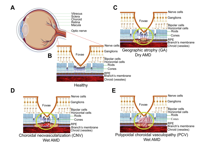
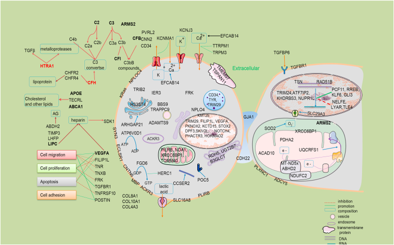
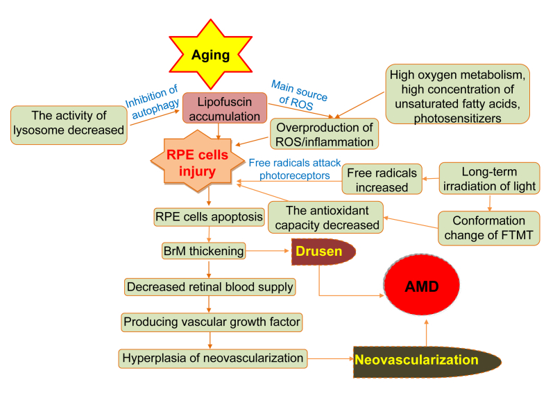
Age-related macular degeneration (AMD) is a complex eye disorder and is the leading cause of incurable blindness worldwide in the elderly. Clinically, AMD initially affects the central area of retina known as the macula and it is classified as early stage to late stage (advanced AMD). The advanced AMD is classified into the nonexudative or atrophic form (dry AMD) and the exudative or neovascular form (wet AMD). More severe vision loss is typically associated with the wet form. Multiple genetic factors, lipid metabolism, oxidative stress and aging, play a role in the etiology of AMD. Dysregulation in genetic to AMD is established to 46%-71% of disease contribution, with CFH and ARMS2/HTRA1 to be the two most notable risk loci among the 103 identified AMD associated loci so far. Chronic cigarette smoking is the most proven consistently risk living habits for AMD. Deep learning algorithm has been developed based on image recognition to distinguish wet AMD and normal macula with high accuracy. Currently, anti-vascular endothelial growth factor (VEGF) therapy is highly effective at treating wet AMD. Several new generation AMD drugs and iPSC-derived RPE cell therapy are in the clinical trial stage and are promising to improve AMD treatment in the near future.

Keywords: Age-related macular degeneration; Diagnosis; Genetics; Mechanism; Target treatment.


Figures

Figure 1
Figure 1
Neovascularization and drusen formation. (A) A cross section of a normal human eye showing the location of the macula. An oval area with a diameter of 1.5 mm near the fovea is known as the macula. (B) It shows the important structure of the retina in the macular area. RPE cells have the function of removing metabolites produced by photoreceptors. The blood vessels in the choroid can transport nutrients and nourish the outer retina. (C) The formation and location of drusen is shown. Drusen formation will lead to retinal tissue atrophy and Bruch membrane calcification rupture, further leading to AMD. (D) This picture describes a choroid polypoid lesion. (E) This picture shows the proliferation of new blood vessels.
Figure 2
Figure 2
The predicted global prevalence of AMD in 2040. TThis figure shows the estimated number of people worldwide with AMD in 2040. Data from REF.10.
Figure 3
Figure 3
Schematic diagram of AMD genetics related genes/proteins. Some of these genes are distributed in the nucleus to participate in the regulation of transcription process, the methylation of histone and so on, some are distributed in the mitochondria to participate in the electron transmission of respiratory chain, and some are distributed in the cytoplasmic matrix and serve as the cytoskeleton used in the cytoskeleton to participate in the regulation of cell life activities. AG: arachidonic acid glyceride.
Figure 4
Figure 4
The formation of AMD during aging. This diagram shows the non-genetic mechanisms of AMD induced by RPE cell senescence, oxidative stress, hemodynamics and so on during aging.
Figure 5
Figure 5
Suggestions on treatment and follow-up of AMD. This is the treatment strategy for AMD. Routine ophthalmic examination and follow-up were the main methods for early and middle stage AMD patients, while appropriate treatment was adopted for advanced AMD patients according to their characteristics.
Figure 6
Figure 6
Schematic diagram of cell therapy and gene therapy for AMD. The fertilized ovum was obtained by in vitro fertilization, and the fertilized ovum developed into a morula and then a blastocyst. The inner cell population of the blastula was proliferated in vitro to obtain embryonic stem cells, which were induced to differentiate into RPE cells and transplanted into the patients’ eyes. In addition, skin fibroblasts from the patient were induced to produce pluripotent stem cells, which differentiated into RPE cells and were transplanted into the patients’ eyes. Gene therapy is performed mainly by injecting AAV vector carrying vVEGF-binding protein into the rsubetinal space of patients.

References

    1. Davis M.D., Gangnon R.E., Lee L.Y., et al. The Age-Related Eye Disease Study severity scale for age-related macular degeneration: AREDS Report No. 17. Arch Ophthalmol. 2005;123(11):1484–1498. - PMC - PubMed
    1. Ferris F.L., Davis M.D., Clemons T.E., et al. A simplified severity scale for age-related macular degeneration: AREDS Report No. 18. Arch Ophthalmol. 2005;123(11):1570–1574. - PMC - PubMed
    1. Lim L.S., Mitchell P., Seddon J.M., Holz F.G., Wong T.Y. Age-related macular degeneration. Lancet. 2012;379(9827):1728–1738. - PubMed
    1. Liew G., Joachim N., Mitchell P., Burlutsky G., Wang J.J. Validating the AREDS simplified severity scale of age-related macular degeneration with 5- and 10-year incident data in a population-based sample. Ophthalmology. 2016;123(9):1874–1878. - PubMed
    1. Fine S.L., Berger J.W., Maguire M.G., Ho A.C. Age-related macular degeneration. N Engl J Med. 2000;342(7):483–492. - PubMed