. 2021 Dec 31;13(1):e12262.
doi: 10.1002/dad2.12262. eCollection 2021.

Predicting disease progression in behavioral variant frontotemporal dementia

Affiliations

Predicting disease progression in behavioral variant frontotemporal dementia

Sarah Anderl-Straub et al. Alzheimers Dement (Amst). .

Abstract

Introduction: The behavioral variant of frontotemporal dementia (bvFTD) is a rare neurodegenerative disease. Reliable predictors of disease progression have not been sufficiently identified. We investigated multivariate magnetic resonance imaging (MRI) biomarker profiles for their predictive value of individual decline.

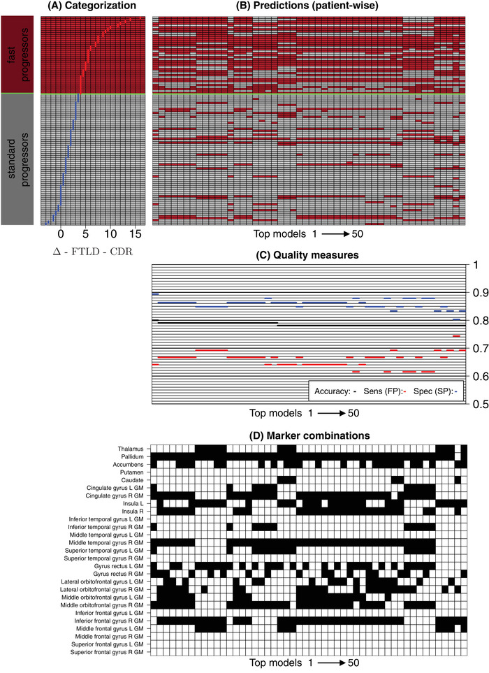
Methods: One hundred five bvFTD patients were recruited from the German frontotemporal lobar degeneration (FTLD) consortium study. After defining two groups ("fast progressors" vs. "slow progressors"), we investigated the predictive value of MR brain volumes for disease progression rates performing exhaustive screenings with multivariate classification models.

Results: We identified areas that predict disease progression rate within 1 year. Prediction measures revealed an overall accuracy of 80% across our 50 top classification models. Especially the pallidum, middle temporal gyrus, inferior frontal gyrus, cingulate gyrus, middle orbitofrontal gyrus, and insula occurred in these models.

Discussion: Based on the revealed marker combinations an individual prognosis seems to be feasible. This might be used in clinical studies on an individualized progression model.

Keywords: behavioral variant frontotemporal dementia; brain volume; classification models; disease progression; frontotemporal dementia; prognosis.


Conflict of interest statement

The authors declare no conflicts of interest.

Figures

FIGURE 1
FIGURE 1
Spearman correlation of brain volumes. The figure provides a heatmap of the pairwise Spearman correlations of the brain volumes (range: [0.41, 0.92]). Additionally, the correlations of the brain values to the Δ‐FTLD‐CDR (range: [–0.35, –0.01]) and the diagnostic groups (range: [–0.37, –0.02]) are shown. The correlations are given numerically (x102) and indicated by the color scheme (green: positive correlation, red: negative correlation). CDR, Clinical Dementia Rating; FTLD, frontotemporal lobar degeneration
FIGURE 2
FIGURE 2
Brain volumes. The figure provides an overview on the declines of brain volumes (ml) of the analyzed gyri for the patient groups slow progressors (blue, Δ‐FTLD‐CDR < 4) and fast progressors (red, Δ‐FTLD‐CDR ≥ 4). Each panel shows a specific gyrus or a pair of gyri. For paired gyri a line indicates the correspondence of the left and right hemisphere. The brain volumes were screened for statistically significant median differences between the group of fast progressors and slow progressors by applying two‐sided Wilcoxon rank‐sum. Significant results were indicated by an asterix (P = 0.05, Bonferroni correction n = 27) . CDR, Clinical Dementia Rating; FTLD, frontotemporal lobar degeneration
FIGURE 3
FIGURE 3
Results of multivariate screening. The figure provides an overview on the results of the screening experiments with multivariate profiles (subsets) of brain volumes. All combinations from one up to 10 gyri (> 1.6 × 107 experiments) were evaluated in LOOCV experiments and ranked according to the accuracy achieved by 1‐NN classifiers. The top 50 marker combinations according to accuracy are shown. A, Overview on the Δ‐FTLD‐CDR scores of the individual patients. The scores are sorted and a green line indicates the border between the groups of fast progressors (red) and slow progressors (gray). The columns of (B‐D) are sorted according to the accuracy of the top 50 marker combinations. The leftmost columns provide the results for the marker combination with the highest accuracy. B, Predictions for the individual patients. The patients (rows) are sorted according to their Δ‐FTLD‐CDR scores (A). The overall accuracies as well as the sensitivities (fast progressors) and specificities (slow progressors) are given in (C). The corresponding brain volume combinations (black) are given in the columns of (D). 1‐NN, nearest neighbor; CDR, Clinical Dementia Rating; FTLD, frontotemporal lobar degeneration; LOOCV, leave‐one‐out cross‐validation

References

    1. Rascovsky K, Hodges JR, Knopman D, et al. Sensitivity of revised diagnostic criteria for the behavioural variant of frontotemporal dementia. Brain. 2011; 134(9): 2456‐2477. 10.1093/brain/awr179 - DOI - PMC - PubMed
    1. Devenney E, Bartley L, Hoon C, et al. Progression in behavioral variant frontotemporal dementia: a longitudinal study. JAMA Neurol. 2015; 72(12): 1501‐1509. - PubMed
    1. Zhutovsky P, Vijverberg EGB, Bruin WB, et al. Individual prediction of behavioral variant frontotemporal dementia development using multivariate pattern analysis of magnetic resonance imaging data. J Alzheimers Dis. 2019; 68(3): 1229‐1241. - PubMed
    1. Staffaroni AM, Ljubenkov PA, Kornak J, et al. Longitudinal multimodal imaging and clinical endpoints for frontotemporal dementia clinical trials. Brain. 2019; 142(2): 443‐459. - PMC - PubMed
    1. Ranasinghe KG, Rankin KP, Pressman PS, et al. Distinct Subtypes of Behavioral Variant Frontotemporal Dementia Based on Patterns of Network Degeneration. JAMA Neurology. 2016; 73(9): 1078. 10.1001/jamaneurol.2016.2016 - DOI - PMC - PubMed