

Comparison of the Retinal and Choroidal Structures in 3 Refractive Groups
- PMID: 35005500
- PMCID: PMC8651023
- DOI: 10.14744/bej.2021.47568
Comparison of the Retinal and Choroidal Structures in 3 Refractive Groups
Abstract
Objectives: This study investigated the retinal layer thickness, choroidal thickness (CT), and retinal nerve fiber layer (RNFL) parameters in 3 refractive groups.
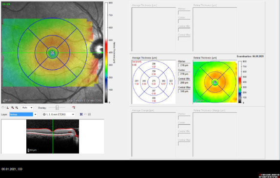
Methods: A total of 201 eyes of 201 subjects were enrolled in this prospective and comparative study. The patients were divided into 3 groups according to refractive status: Group 1 consisted of 60 eyes of myopic subjects, Group 2 comprised 72 eyes of emmetropic subjects, and 69 eyes of hyperopic subjects were categorized as Group 3. The retinal layer thickness, CT, and RNFL parameters were measured using optical coherence tomography and compared between groups.
Results: The mean age of the patients was 22.33±10.11 years in Group 1, 21.55±8.3 years in Group 2, and 23.73±11.08 years in Group 3 (p=0.741). Group 1 consisted of 34 women and 26 men, Group 2 contained 44 women and 28 men, and Group 3 was made up of 45 women and 24 men (p=0.124). The mean spherical equivalent value was -6.16±2.01 D in Group 1, 0.13±0.5 D in Group 2, and 5.48±1.32 D in Group 3 (p<0.001). The RNFL and macular thickness values were lower in the myopic patients compared with those of the other groups (p<0.05). The CT measurement was lower in the myopic patients and higher in the hyperopic patients compared with the emmetropic patients (p<0.05).
Conclusion: The myopic patients had a lower CT and RNFL thickness measurement than the emmetropic and hyperopic patients, whereas the hyperopic patients had a higher CT than the other patient types.
Keywords: Choroidal thickness; emmetropia; hyperopia; myopia; retinal nerve fiber layer.
Copyright: © 2021 by Beyoglu Eye Training and Research Hospital.
Conflict of interest statement
Conflict of Interest: None declared.
Figures

References
-
- Imamura Y, Fujiwara T, Margolis R, Spaide RF. Enhanced depth imaging optical coherence tomography of the choroid in central serous chorioretinopathy. Retina. 2009;29:1469–73. - PubMed
-
- Spaide RF, Koizumi H, Pozzoni MC. Enhanced depth imaging spectral-domain optical coherence tomography. Am J Ophthalmol. 2008;146:496–500. - PubMed
-
- Kim SW, Oh J, Kwon SS, Yoo J, Huh K. Comparison of choroidal thickness among patients with healthy eyes, early age-related maculopathy, neovascular age-related macular degeneration, central serous chorioretinopathy, and polypoidal choroidal vasculopathy. Retina. 2011;31:1904–11. - PubMed
LinkOut - more resources
Full Text Sources