

2D Analysis of Gold Weight Implantation Surgery Results in Paralytic Lagophthalmos
- PMID: 35005516
- PMCID: PMC8697052
- DOI: 10.14744/bej.2021.95866
2D Analysis of Gold Weight Implantation Surgery Results in Paralytic Lagophthalmos
Abstract
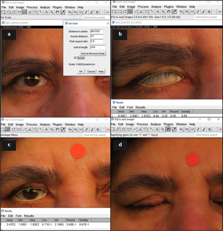
Objectives: Gold weight implantation in the upper eyelid is a frequently performed treatment for paralytic lagophthalmos to prevent corneal exposure. A margin reflex distance of -1 and -2 (MRD1, MRD2), the palpebral fissure height (PFH), and the vertical lagophthalmos (LV) are 1-dimensional (1D) measurements used in follow-up. Because the exposure area is 2-dimensional (2D), this study was designed to investigate the results using both 1D and 2D analysis.
Methods: Ten patients who underwent pretarsal suborbicularis oculi gold weight implantation were included in the study. Photographs were taken with a digital camera and the images were analyzed using ImageJ software (US National Institutes of Health, Bethesda, MD, USA). The lagophthalmos area (LA) and ocular surface area (OSA) were measured in 2D in addition to the MRD1, MRD2, PFH, LV. Preoperative and postoperative values were compared using the Wilcoxon signed-rank test. Associations between parameters were evaluated using Spearman's correlation analysis.
Results: The mean age of the patients (7 male, 3 female) was 39.6±16.4 years (range: 14-60 years). The mean implant weight was 1.46 g (0.8-1.6 g). There were significant reductions in the MRD1, MRD2, PFH, OSA, LV, and LA values after surgery (p<0.05). The weight of the gold implant had a strong correlation with the PFH, OSA, MRD1, and MRD2, but not the LV or LA, preoperatively. The OSA was strongly correlated with the MRD1, PFH, and the implant weight, but not the MRD2. The LA was strongly correlated with the LV, preoperatively. In the postoperative period, the OSA was strongly correlated with the PFH and the MRD2 but not the MRD1, while the LA was strongly correlated with the LV, MRD1, and the PFH.
Conclusion: It is easy to obtain 2D measurements using digital image analysis software, and they proved to be accurate and correlated strongly with 1D measurements. The OSA and LA measurements were significantly lower following upper eyelid gold weight implantation. The PFH and LV were compatible with the OSA and LA, preoperatively.
Keywords: Gold weight; ImageJ; image analysis; paralytic lagophthalmos.
Copyright © 2021 by Beyoglu Eye Training and Research Hospital.
Figures
References
-
- Vásquez LM, Medel R. Lagophthalmos after facial palsy: current therapeutic options. Ophthalmic Res. 2014;52:165–9. - PubMed
-
- Mancini R, Taban M, Lowinger A, Pariseau B, Leyngold AR, Anderson RL. Use of hyaluronic acid gel in the management of paralytic lagophthalmos: the hyaluronic acid gel ‘gold weight’. Ophthal Plast Reconstr Surg. 2009;25:23–6. - PubMed
-
- Rofagha S, Seiff SR. Long-term results for the use of gold eyelid load weights in the management of facial paralysis. Plast Reconstr Surg. 2010;125:142–9. - PubMed
-
- Hassan AS, Frueh BR, Elner VM. Müllerectomy for upper eyelid retraction and lagophthalmos due to facial nerve palsy. Arch Ophthalmol. 2005;123:1221–5. - PubMed
-
- Guillou-Jamard MR, Labbé D, Bardot J, Benateau H. Paul Tessier's technique in the treatment of paralytic lagophthalmos by lengthening of the levator muscle: evaluation of 29 cases. Ann Plast Surg. 2011;67:S31–5. - PubMed
LinkOut - more resources
Full Text Sources