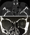
Delayed Management of an Orbital Floor Blow-out Fracture
- PMID: 35005524
- PMCID: PMC8697045
- DOI: 10.14744/bej.2021.94834
Delayed Management of an Orbital Floor Blow-out Fracture
Abstract
A bony fracture in the orbital floor, the most common site, can lead to tissue herniation, enophthalmos, hypoglobus, or strabismic diplopia. Several surgical approaches for repair have been described in the literature. This report is a description of an illustrative case and a brief summary of the literature related to the transconjunctival approach to orbital floor fracture repair as performed by ophthalmologists. A 19-year-old female patient had fallen from a 5-meter-high fence and sustained panfacial fractures, including both orbits and the surrounding sinuses. An acute repair was performed by a maxillofacial team to stabilize the facial structure . Following neurosurgical stabilization, she was referred to ophthalmology with pronounced hypoglobus and enophthalmos, diplopia, relative afferent pupillary defect, and a slightly pale right optic nerve head. Surgery was performed under general anesthesia using the transconjunctival approach and an alloplastic implant. This approach was effective, providing excellent exposure while reducing the risks of lower eyelid retraction and surgical scars associated with the transcutaneous approach.
Keywords: Enophthalmos; hypoglobus; orbital fracture; transconjunctival approach.
Copyright © 2021 by Beyoglu Eye Training and Research Hospital.
Figures


Similar articles
-
Intact Periorbita Can Prevent Post-Traumatic Enophthalmos Following a Large Orbital Blow-Out Fracture.Craniomaxillofac Trauma Reconstr. 2020 Mar;13(1):49-52. doi: 10.1177/1943387520903545. Epub 2020 Mar 23. Craniomaxillofac Trauma Reconstr. 2020. PMID: 32642032 Free PMC article.
-
Clinical recommendations for repair of isolated orbital floor fractures: an evidence-based analysis.Ophthalmology. 2002 Jul;109(7):1207-10; discussion 1210-1; quiz 1212-3. doi: 10.1016/s0161-6420(02)01057-6. Ophthalmology. 2002. PMID: 12093637 Review.
-
Delayed immediate surgery for orbital floor fractures: Less can be more.Can J Plast Surg. 2011 Winter;19(4):125-8. Can J Plast Surg. 2011. PMID: 23204882 Free PMC article.
-
Endoscopic repair of orbital floor fractures.Otolaryngol Clin North Am. 2007 Apr;40(2):319-28. doi: 10.1016/j.otc.2006.11.007. Otolaryngol Clin North Am. 2007. PMID: 17383511
-
Endoscopic repair of orbital floor fractures.Facial Plast Surg Clin North Am. 2006 Feb;14(1):11-6. doi: 10.1016/j.fsc.2005.11.001. Facial Plast Surg Clin North Am. 2006. PMID: 16466978 Review.
Cited by
-
Subcutaneous emphysema and pneumomediastinum following orbital blowout pathological fracture in a cat with nasal lymphoma: a case report.BMC Vet Res. 2023 Sep 15;19(1):161. doi: 10.1186/s12917-023-03722-0. BMC Vet Res. 2023. PMID: 37715215 Free PMC article.
-
Delayed Orbital Floor Reconstruction Using Mirroring Technique and Patient-Specific Implants: Proof of Concept.J Pers Med. 2024 Apr 26;14(5):459. doi: 10.3390/jpm14050459. J Pers Med. 2024. PMID: 38793041 Free PMC article.
References
-
- Shin JW, Lim JS, Yoo G, Byeon JH. An analysis of pure blowout fractures and associated ocular symptoms. J Craniofac Surg. 2013;24:703–7. - PubMed
-
- Koenen L, Waseem M. Orbital Floor Fracture. StatPearls. Treasure Island, FL: StatPearls Publishing; 2020. Available at: https://www.ncbi.nlm.nih.gov/books/NBK534825/. Accessed May 3, 2021. - PubMed
-
- Burnstine MA. Clinical recommendations for repair of orbital facial fractures. Curr Opin Ophthalmol. 2003;14:236–40. - PubMed
Publication types
LinkOut - more resources
Full Text Sources
