. 2021 Dec 9;25(1):103588.
doi: 10.1016/j.isci.2021.103588. eCollection 2022 Jan 21.

IL-27 induces IFN/STAT1-dependent genes and enhances function of TIGIT+ HIVGag-specific T cells

Affiliations

IL-27 induces IFN/STAT1-dependent genes and enhances function of TIGIT+ HIVGag-specific T cells

Jie Cheng et al. iScience. .

Abstract

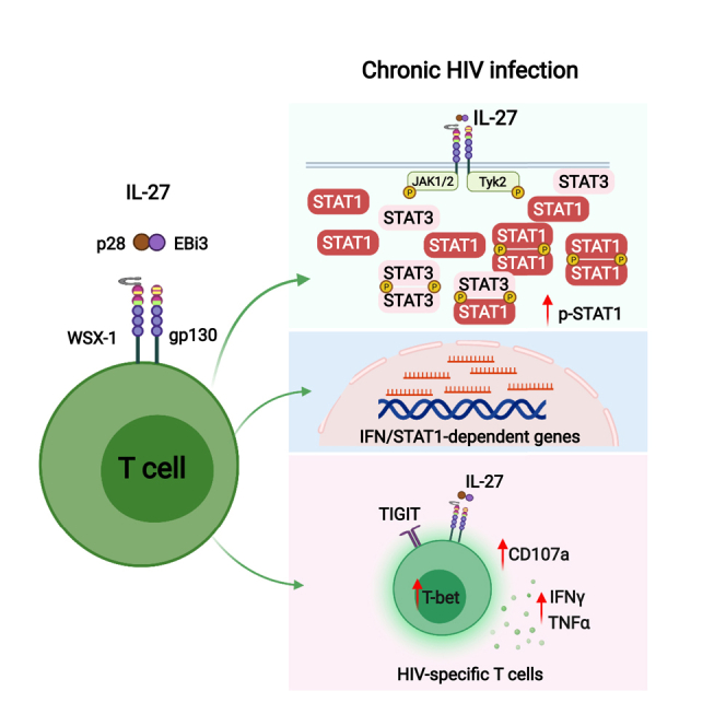
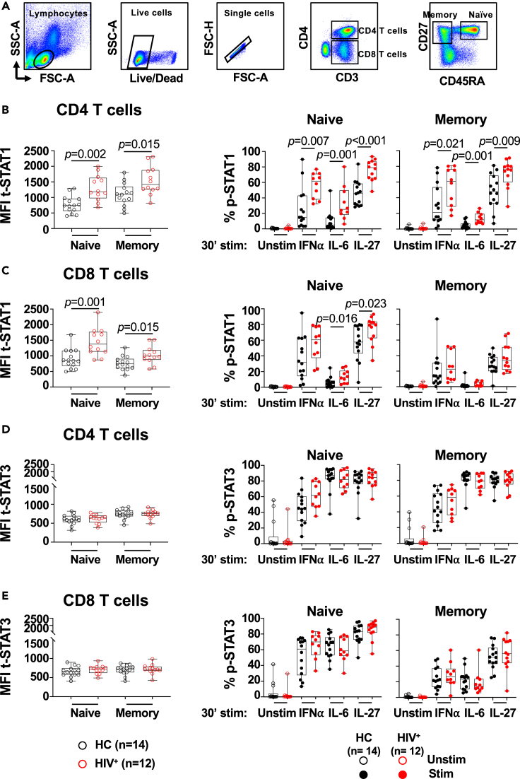
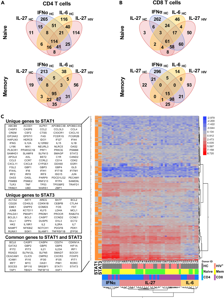
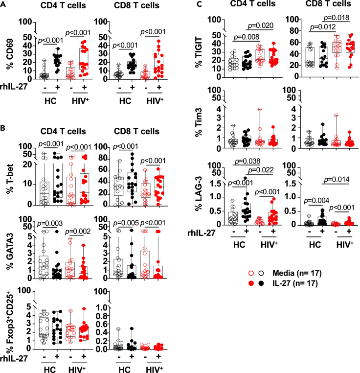
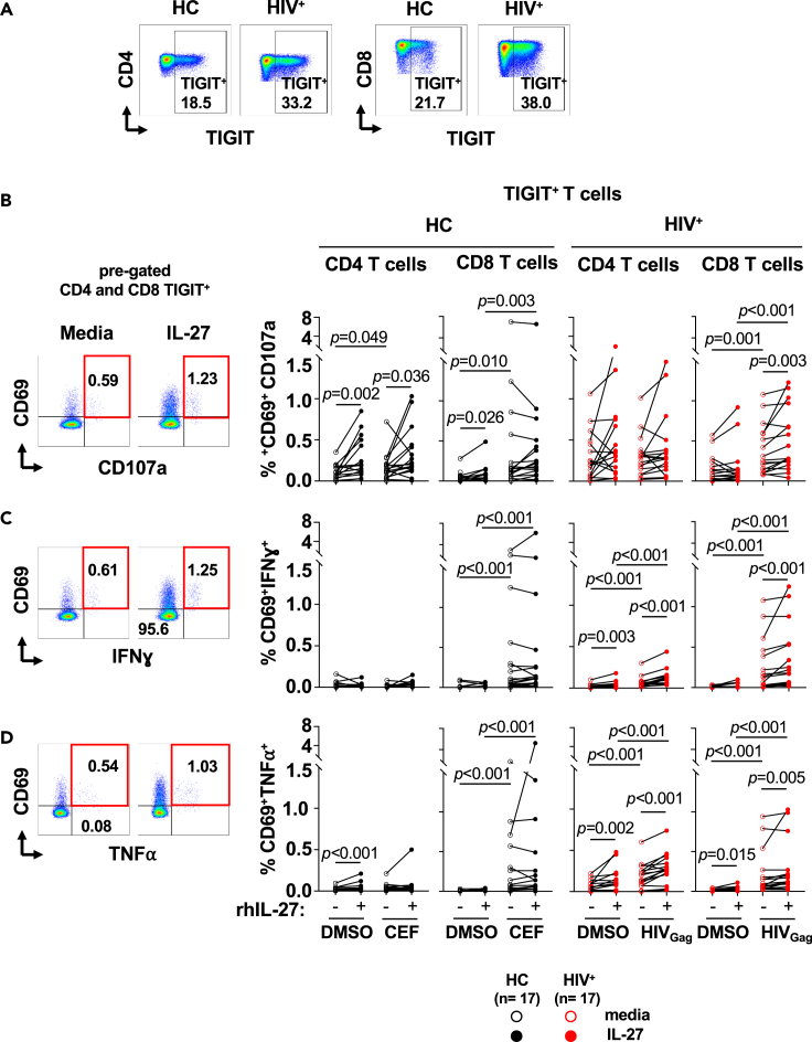
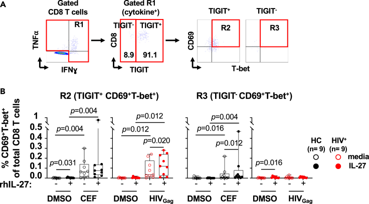
HIV-specific T cells have diminished effector function and fail to control/eliminate the virus. IL-27, a member of the IL-6/IL-12 cytokine superfamily has been shown to inhibit HIV replication. However, whether or not IL-27 can enhance HIV-specific T cell function is largely unknown. In the present manuscript, we investigated the role of IL-27 signaling in human T cells by evaluating the global transcriptional changes related to the function of HIV-specific T cells. We found that T cells from people living with HIV (PLWH), expressed higher levels of STAT1 leading to enhanced STAT1 activation upon IL-27 stimulation. Observed IL-27 induced transcriptional changes were associated with IFN/STAT1-dependent pathways in CD4 and CD8 T cells. Importantly, IL-27 dependent modulation of T-bet expression promoted IFNγ secretion by TIGIT+HIVGag-specific T cells. This new immunomodulatory effect of IL-27 on HIV-specific T cell function suggests its potential therapeutic use in cure strategies.

Keywords: Components of the immune system; Immune response; Transcriptomics.


Conflict of interest statement

The authors declare no competing interests.

Figures

None
Graphical abstract
Figure 1
Figure 1
In vitro IL-27 stimulation leads to enhanced STAT1 activation in naive and memory phenotype T cells from PLWH PBMCs from PLWH (HIV+, n = 12) and healthy controls (HC, n = 14) were thaw, rested overnight and stimulated with 25 ng/mL of rhIL-27, 100 U/mL of rhIFN⍺, and 100 ng/mL of rhIL-6, for 30 min. (A) Gating strategy of naive (CD45RA+CD27+) and memory (CD45RACD27+) phenotype CD4 and CD8 T cells. Median Fluorescence Intensity (MFI) of intracellular expression of t-STAT1 and frequency of phosphorylated STAT1 (p-STAT1) in: (B) naive and memory phenotype CD4 T cell subsets; (C) naive and memory phenotype CD8 T cell subsets. Median Fluorescence Intensity (MFI) of intracellular expression of t-STAT3 and frequency of phosphorylated STAT3 (p-STAT3) in: (D) naive and memory phenotype CD4 T cell subsets; and (E) naive and memory phenotype CD8 T cell subsets. The graph is represented by box and whisker showing the median value with first and third quartiles in the box, with whiskers extending to the minimum and maximum values. Comparisons between the study groups PLWH and healthy controls were performed using Mann–Whitney test. p value <0.05 was considered significant.
Figure 2
Figure 2
IL-27 signaling induced a cluster of STAT1-dependent genes RNAseq analysis of sorted naive (CD45RA+CD27+) and memory (CD45RACD27+) T cells from PLWH (HIV+, n = 5) and healthy controls (HC, n = 3) stimulated in vitro with 100 ng/mL of rhIL-27, and cells from healthy controls stimulated with 100 U/mL of rhIFN⍺ and 100 ng/mL of rhIL-6 for 90 min as controls. (A) Venn diagrams represent overlapped differentially expressed gene transcripts (DEGs) regulated by IL-27 in CD4 T cell subsets from PLWH and HC compared to the DEGs induced by IFN⍺ and IL-6 stimulation in T cells from healthy controls. (B) Venn diagrams represent overlapped DEGs regulated by IL-27 in CD8 T cell subsets from PLWH and HC compared to the DEGs induced by IFN⍺ and IL-6 stimulation in T cell from healthy controls. (C) Ingenuity Pathway Analysis (IPA) was used to subset the RNA-seq data to include only target genes predicted by IPA to be regulated via STAT1 and/or STAT3 activity, as indicated in the left column, orange for genes downstream, gray for genes not downstream of the corresponding upstream regulator. The 2-way clustered heatmap of gene expression (log2 of stimulated/unstimulated ratio) for IL-27, IFNα and IL-6 stimulated T cell subsets. The color bar indicates: HC group (gray), HIV group (pink), CD4 T cells (blue), CD8 T cells (purple), naive subset (green), Memory (Mem, yellow). The lists (DEGs) were selected based on >2-fold change (| Log2 FC | > 1) and FDR <0.05.
Figure 3
Figure 3
In vitro IL-27 stimulation increased expression of CD69 and T-bet in CD4 and CD8 T cells PBMCs from PLWH (HIV+, n = 17) and healthy controls (HC, n = 17) were cultured in the absence (opened symbols) or presence (closed symbols) of rhIL-27 (100 ng/mL) overnight. Flow cytometry analysis was performed for the expression of: (A) CD69; (B) T-bet, GATA3, and Foxp3+CD25+; (C) TIGIT, Tim3 and LAG3 in CD4 and CD8 T cells. Expressions of the markers are expressed as frequencies of the CD4 and CD8 T cells. The graphs are represented by box and whisker showing the median value with first and third quartiles in the box, with whiskers extending to the minimum and maximum values. Comparisons between the media and IL-27 stimulation were performed by non-parametric Wilcoxon test. Comparisons between the groups were performed by non-parametric Mann–Whitney test. p value <0.05 was considered significant.
Figure 4
Figure 4
IL-27 enhances cytokine secretion of TIGIT+ HIVGag-specific T cells PBMCs from PLWH (HIV+, n = 17) were cultured in the absence (red opened symbols) or presence (red closed symbols) of rhIL-27 (100 ng/mL) overnight follow by stimulation with HIVGag peptide pool. DMSO was used as control. PBMCs from and healthy controls (HC, n = 17) were stimulated in the absence (black opened symbols) or presence (black closed symbols) of rhIL-27 (100 ng/mL) overnight follow by stimulation with CEF (CMV, EBV and Influenza pool of peptides), and DMSO as control. Secretion of cytokines by TIGIT+ T cells was analyzed by flow cytometry. (A) Flow cytometry gating strategy for TIGIT+ CD4 and CD8 T cells. TIGIT+ CD4 and CD8 T cells were analyzed for expression of: (B) CD69+CD107a+; (C) CD69+IFNɣ+; and (D) CD69+TNF⍺+. The graphs are represented by box and whisker showing the median value with first and third quartiles in the box, with whiskers extending to the minimum and maximum values of the frequencies of T cells secreting cytokines. Comparisons between the culture conditions were performed by Wilcoxon test. Comparisons between PLWH and healthy controls were performed by Mann–Whitney test. p value <0.05 was considered significant.
Figure 5
Figure 5
Enhanced TIGIT+HIVGag-specific CD8 T cell function is driven by IL-27 dependent upregulation of T-bet PBMCs from PLWH (HIV+, n = 9) were cultured in the absence (red opened symbols) or presence (red closed symbols) of rhIL-27 (100 ng/mL) overnight follow by stimulation with HIVGag peptide pool. DMSO was used as control. PBMCs from and healthy controls (HC, n = 9) were cultured in the absence (black opened symbols) or presence (black closed symbols) of rhIL-27 (100 ng/mL) overnight follow by stimulation with were stimulated with CEF (CMV, EBV and Influenza pool of peptides), and DMSO was used as control. IL-27 upregulation of T-bet and CD69 was measured by flow cytometry. (A) Gating strategy for TIGIT+and TIGITvirus-specific T cells secreting IFNγ+TNFα+. (B) Expression of CD69+T-bet+ in cytokine secreting CD8 T cells. TIGIT+CD69+T-bet+ or TIGIT CD69+T-bet+ cytokine secreting T cells is expressed as frequency of total CD8 T cells. The graph is represented by box and whisker showing the median value with first and third quartiles in the box, with whiskers extending to the minimum and maximum values. Comparisons between culture conditions were performed using non-parametric Wilcoxon test. p value <0.05 was considered significant.

References

    1. Batten M., Kljavin N.M., Li J., Walter M.J., de Sauvage F.J., Ghilardi N. Cutting edge: IL-27 is a potent inducer of IL-10 but not FoxP3 in murine T cells. J. Immunol. 2008;180:2752–2756. - PubMed
    1. Betts M.R., Nason M.C., West S.M., De Rosa S.C., Migueles S.A., Abraham J., Lederman M.M., Benito J.M., Goepfert P.A., Connors M., et al. HIV nonprogressors preferentially maintain highly functional HIV-specific CD8+ T cells. Blood. 2006;107:4781–4789. - PMC - PubMed
    1. Buggert M., Frederiksen J., Lund O., Betts M.R., Biague A., Nielsen M., Tauriainen J., Norrgren H., Medstrand P., Karlsson A.C., et al. CD4+ T cells with an activated and exhausted phenotype distinguish immunodeficiency during aviremic HIV-2 infection. AIDS. 2016;30:2415–2426. - PMC - PubMed
    1. Buggert M., Nguyen S., McLane L.M., Steblyanko M., Anikeeva N., Paquin-Proulx D., Del Rio Estrada P.M., Ablanedo-Terrazas Y., Noyan K., Reuter M.A., et al. Limited immune surveillance in lymphoid tissue by cytolytic CD4+ T cells during health and HIV disease. PLoS Pathog. 2018;14:e1006973. - PMC - PubMed
    1. Catalfamo M., Di Mascio M., Hu Z., Srinivasula S., Thaker V., Adelsberger J., Rupert A., Baseler M., Tagaya Y., Roby G., et al. HIV infection-associated immune activation occurs by two distinct pathways that differentially affect CD4 and CD8 T cells. Proc. Natl. Acad. Sci. U S A. 2008;105:19851–19856. - PMC - PubMed