. 2021 Dec:8:179-201.
doi: 10.3114/fuse.2021.08.14. Epub 2021 Nov 29.

Anchoring the species Rhizophagus intraradices (formerly Glomus intraradices)

Affiliations

Anchoring the species Rhizophagus intraradices (formerly Glomus intraradices)

C Walker et al. Fungal Syst Evol. 2021 Dec.

Abstract

The nomenclatural type material of Rhizophagus intraradices (basionym Glomus intraradices) was originally described from a trap pot culture established with root fragments, subcultures of which later became registered in the INVAM culture collection as FL 208. Subcultures of FL 208 (designated as strain ATT 4) and a new strain, independently isolated from the type location (ATT 1102), were established as both pot cultures with soil-like substrate and in vitro root organ culture. Long-term sampling of these cultures shows spores of the species to have considerable morphological plasticity, not described in the original description. Size, shape and other features of the spores were much more variable than indicated in the protologue. Phylogenetic analyses confirmed earlier published evidence that sequences from all R. intraradices cultures formed a monophyletic clade, well separated from, and not representing a sister clade to, R. irregularis. Moreover, new phylogenetic analyses show that Rhizoglomus venetianum and R. irregularis are synonymous. The morphological characters used to separate these species exemplify the difficulties in species recognition due to the high phenotypic plasticity in the genus Rhizophagus. Rhizophagus intraradices is morphologically re-described, an epitype is designated from a single-spore isolate derived from ATT 4, and R. venetianum is synonymised with R. irregularis.

Keywords: Glomeromycota; arbuscular mycorrhizal fungi; molecular phylogeny; phenotypic plasticity; species definition.


Figures

Fig. 1.
Fig. 1.
Guidance for measuring spores of glomeromycotan fungi, including standard spheroid descriptions (adapted from Kirk et al. 2010) and some common different shape outlines.
Fig. 2.
Fig. 2.
Phylogenetic maximum likelihood tree of Rhizophagus species and isolates ATT 4 (FL 208) and ATT 1102 of R. intraradices. For the completely resolved and annotated tree see Fig. S2. A. Characterised Rhizophagus species, with Sclerocystis as outgroup. Size of triangles represent the sequence numbers (vertically) and distances (horizontally). B. Details of R. intraradices, showing that descendants (ATT 4-38, ATT 4-41, ATT 4-64) of the ex-type culture FL 208, including (red typeface) the culture from which the epitype was taken (MUCL 52327 = ATT 4-83) and (blue typeface) the strain (MUCL 49410 = ATT 1102-12) newly isolated from the type locality cluster in the same monophyletic clade. C. Details of R. irregularis, showing that “Rhizoglomus venetianum” (red typeface) represents one subtype of the DNA sequence variants of R. irregularis; sequence variants annotated ‘RIRrrna##’ are from a genome sequencing project (Maeda et al. 2018).
Fig. 3.
Fig. 3.
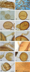
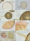
Micromorphology of the holotype specimen of Rhizophagus intraradices (OSC 40255, 5 May 1981). A. Intraradical spores. B. With extra-radical spores singly and in clusters. C. Thin-walled spores extruded from a crushed root. D. Globose spore with subtending hypha (SH) detached close to the spore. E. Broadly ellipsoid spore. F. ‘Pip-shaped’ spore. G. Parallel-sided SH. H. Recurved SH. I. Recurved angular SH with lateral protrusion. J. SH with tubaeform flare. K. Crushed spore showing separation of wall components. L. Outer wall with thickening by bacterial colonies. M. Wall components separating on crushing. N. Bacterial colonies giving an impression of ornamentation. Scale bars: A = 250 μm; B = 1 mm; C–F, K–M = 50 μm; G–J, N = 25 μm.
Fig. 4.
Fig. 4.
Rhizophagus intraradices strain re-isolated from type locality in 1974. A. Intraradical spores. B. With extra-radical spores singly and in clusters. C. Thin-walled spores extruded from a crushed root. D. Globose spore with subtending hypha (SH) detached close to the spore. E. Broadly ellipsoid spore. F. ‘Pip-shaped’ spore. G. Parallel-sided SH. H. Recurved SH. I. Recurved angular SH with lateral protrusion. J. SH with tubaeform flare. K. Crushed spore showing separation of wall components. L. Outer wall with thickening by bacterial colonies. M. Wall components separating on crushing. N. Bacterial colonies giving an impression of ornamentation. Scale bars: A = 250 μm; B = 1 mm; C–M = 50 μm; N = 25 μm.
Fig. 5.
Fig. 5.
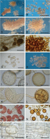
Rhizophagus intraradices from pot cultures. A. Intra- and extra-radical spores. B. Extracted spores that were formed singly. C. Spores bursting through the root cortex and epidermis. D. Aggregation (fascicle) of pale extraradical spores. E. Spores formed in the root cortex. F. Fascicle of darkly coloured spores. G. Loose cluster of extraradical spores. H. Small, dense clusters of spores. I. Almost colourless young spore with expanding outermost wall component. J. Older spore developing pigmentation and with colourless outer component. K. Crushed spore showing wall components 2 & 3, having lost the evanescent outermost component. L. Darkly coloured spore retaining colourless outer component. M. Cluster of young spores with red reaction to Melzer’s reagent. N. Cluster of old spores lacking a reaction to Melzer’s reagent. O. Intraradical spore showing cap-like distal thickening of wall component 2. P. Misshapen (irregular) intraradical spore. Scale bars: A = 1 mm; B–D, H = 500 μm; E, G = 150 μm; F = 250 μm; I, K, M, P = 100 μm; N = 200 μm; J, L, O = 50 μm.
Fig. 6.
Fig. 6.
Rhizophagus intraradices from root organ cultures. A. Intra- and extra-radical spores. B. Extracted spores that were formed singly. C. Spores bursting through the root cortex and epidermis. D. Aggregation (fascicle) of pale extraradical spores. E. Spores formed in the root cortex. F. Fascicle of darkly coloured spores. G. Loose cluster of extraradical spores. H. Small, dense clusters of spores. I. Almost colourless young spore with expanding outermost wall component. J. Older spore developing pigmentation and with colourless outer component. K. Crushed spore showing wall components 2 & 3, having lost the evanescent outermost component. L. Darkly coloured spore retaining colourless outer component. M. Cluster of young spores with red reaction to Melzer’s reagent. N. Cluster of old spores lacking a reaction to Melzer’s reagent. O. Intraradical spore showing cap-like distal thickening of wall component 2. P. Misshapen (irregular) intraradical spore. Scale bars: A = 1 mm; B–D, H = 500 μm; E = 150 μm; F, G = 250 μm; I–L, O, P = 50 μm; M, N = 200 μm.
Fig. 7.
Fig. 7.
Some examples of the many sizes, colours and shapes of spores of Rhizophagus intraradices (basionym Glomus intraradices). Scale bars: A–N, P, R, S, U, Y = 100 μm; O = 50 μm; Q, T, V, W, X = 150 μm.
Fig. 8.
Fig. 8.
Examples of the convoluted and irregular shapes found amongst pot cultures, but rarely in root organ cultures, of Rhizophagus intraradices spores. Scale bars: A = 250 μm; B, G = 200 μm; C–F, J = 100 μm; H–I = 150 μm.
Fig. 9.
Fig. 9.
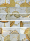
Some of the variation among subtending hyphae of Rhizophagus intraradices. A. Parallel-sided, slightly flared proximally. B. Slightly recurved and narrowed at spore base. C. Gradually narrowing towards spore. D. Convoluted, branched. E. Short branch, narrowing towards spore. F. Funnel-shaped. G. Recurved, with tubaeform flare. H. Recurved, angular, flared. I. Laterally budded (lacking stalk). J. Swollen distally, tapering proximally. K. Recurved, angular with lateral peg. L. Flared, tapering slightly distally. M. Flared, tapering slightly proximally. N. Sharply recurved, expanded towards the spore base. O. Subangular, swollen and flared at the spore base. P. Thickened and constricted proximally. Q. Short branched, tubaeform. R. Tapering proximally, slightly flared, with septal occlusion. Scale bars = 20 μm.
Fig. 10.
Fig. 10.
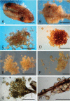
Rhizophagus intraradices clusters of spores. A. Spores occupying an empty Plantago lanceolata seed. B. A dense cluster of spores amongst fine mycelium surrounding a root fragment. C–F. Spores in clusters of varying density showing colour variation. G. Cluster of spores bursting through the cortex of a decaying root. H. Spores clustering around the surface of a decaying root. Scale bars: A = 1 mm; B–H = 500 μm.
Fig. 11.
Fig. 11.
Main spore characteristics of Rhizophagus intraradices. A. Immature spore from ROC showing expansion and apparent layering of the outer component. B. Semi-mature spore with outer component still intact and pigmentation of main structural wall. C. Mature spore from root organ culture (ROC). D. Detail of the wall structure of the spore in C, showing three wall components (1–3): 1, evanescent before degradation; 2, yellow, finely laminate pale yellow; 3, coarsely laminated dark brown. E, F. Crushed spores in polyvinyl alcohol lacto-glycerol with Melzer’s reagent (PVLG-M) showing pale pink reaction of the evanescent wall component. G. Different reactions to PVLG-M; right no reaction, left outer component pink. H. Parallel-sided subtending hypha with slight tubaeform flare and proximal wall thickening. Scale bars: A, B, G = 50 μm; C, E, F = 100 μm; D, H = 25 μm.
Fig. 12.
Fig. 12.
Rhizophagus intraradices: distribution of spore shapes over all treatments (n = 2 673). The ‘other shapes’ section includes all those occurring with a frequency < 2 %.
Fig. 13.
Fig. 13.
Rhizophagus intraradices: distribution (percentage of measured spores) of the spore shapes between those from ATT 4 (Panel A) (n = 1 924) and ATT 1102 (Panel B) (n = 771). Other shapes include those occurring with a frequency < 2 %.
Fig. 14.
Fig. 14.
Rhizophagus intraradices; distribution (percentage of measured spores) of the spore shapes between extraradical spores (Panel A) (n = 1 841) and intraradical spores (Panel B) (n = 737). Other shapes include those occurring with a frequency < 2 %.
Fig. 15.
Fig. 15.
Rhizophagus intraradices: distribution (percentage of measured spores) of the spore shapes between (Panel A) pot cultures (n = 1 559) and (Panel B) root organ cultures (n = 899). Other shapes include those occurring with a frequency < 2 %.
Fig. 16.
Fig. 16.
Rhizophagus intraradices distribution of the four most abundant spore shapes (globose, subglobose, broadly ellipsoid, and ellipsoid) in relation to their host plant. For each panel, the culture method (Pot Culture, PC or Root Organ Culture, ROC) and the number of spores measured are given. Values are given as a percentage of the total spores measured. When a percentage is very low and the colour barely visible (i.e., < 1 %), the colour legend is indicated next to the number.
Fig. 17.
Fig. 17.
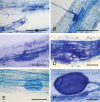
Rhizophagus intraradices mycorrhiza with Plantago lanceolata as host, cleared with KOH and stained with 0.02 % methyl blue in 0.1 M HCl. A. Crushed fine root showing arbuscular mycorrhiza and intraradical spores. B. Appressorium at entry point into root cortex. C. Finely branched arbuscule. D. Entry point showing hyphal coils in outer cortical cells and a thin-walled structure, either a vesicle or an immature spore. E. Thin-walled structure in the cortex which could be interpreted as a vesicle, but may be an intraradical spore in the earliest development stage. F. Thick-walled mature intraradical spore. Scale bars: A = 250 μm, B = 125 μm, C–F = 50 μm.

Similar articles

Cited by

References

    1. Alaux P-L, Mison C, Senés-Guerrero, et al. (2021). Diversity and species composition of arbuscular mycorrhizal fungi across maize fields in the southern part of Belgium. Mycorrhiza 31: 265–272. - PubMed
    1. Anon (1969). Royal Botanic Garden Edinburgh, Flora of the British Fungi Colour Identification Chart. HMSO, Edinburgh, UK.
    1. Anon (1990). Munsell Soil Color Charts 1990 edition revised. Munsell Color Macbeth Division of Kollmorgen Instruments Corp., Maryland, USA.
    1. Błaszkowski J, Czerniawska B, Wubet T, et al. (2008). Glomus irregulare, a new arbuscular mycorrhizal fungus in the Glomeromycota. Mycotaxon 106: 247–267.
    1. Butler EJ. (1939). The occurrence and systematic position of the vesicular-arbuscular type of mycorrhizal fungi. Transactions of the British Mycological Society 22: 274–301

LinkOut - more resources