. 2021 Dec 23:13:100194.
doi: 10.1016/j.mtbio.2021.100194. eCollection 2022 Jan.

Immunomodulatory functions of human mesenchymal stromal cells are enhanced when cultured on HEP/COL multilayers supplemented with interferon-gamma

Affiliations

Immunomodulatory functions of human mesenchymal stromal cells are enhanced when cultured on HEP/COL multilayers supplemented with interferon-gamma

Mahsa Haseli et al. Mater Today Bio. .

Abstract

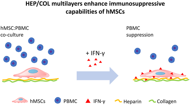
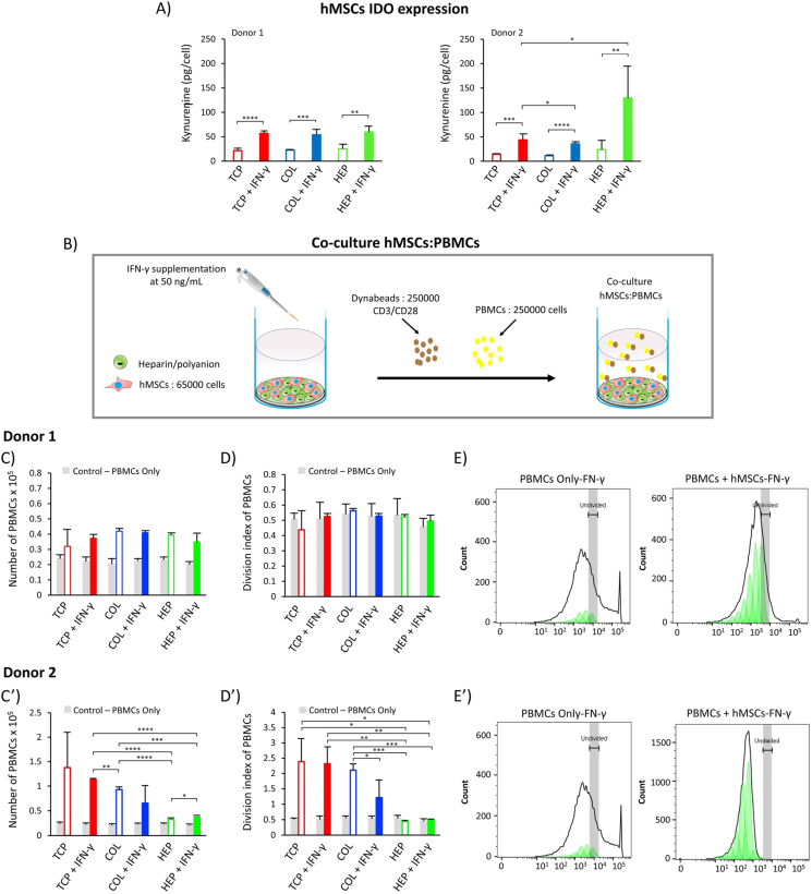
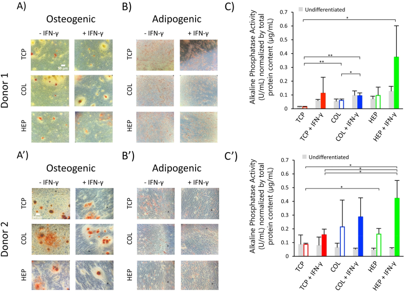
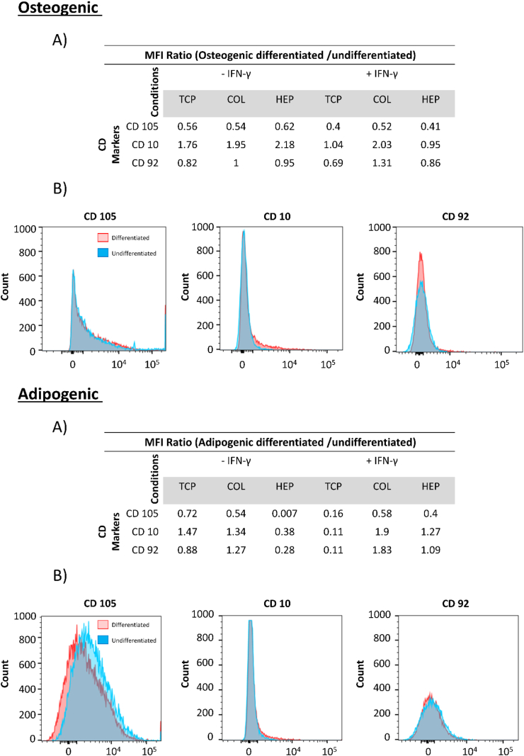
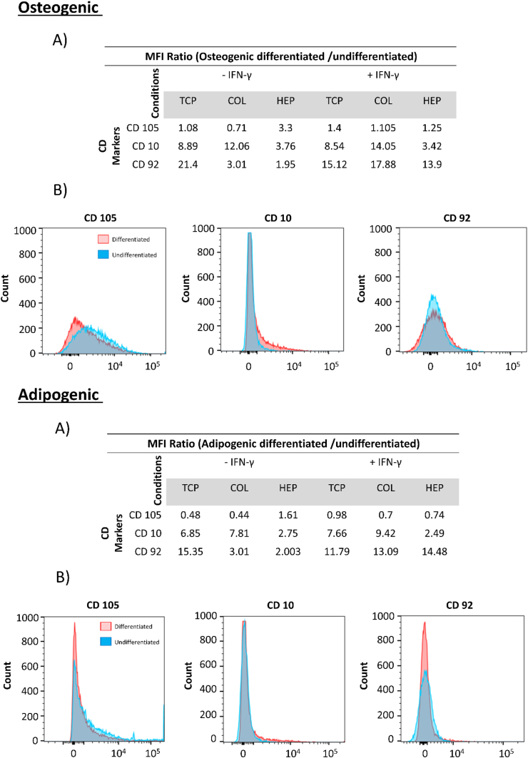
Human mesenchymal stromal cells (hMSCs) are multipotent cells that have been proposed for cell therapies due to their immunosuppressive capacity that can be enhanced in the presence of interferon-gamma (IFN-γ). In this study, multilayers of heparin (HEP) and collagen (COL) (HEP/COL) were used as a bioactive surface to enhance the immunomodulatory activity of hMSCs using soluble IFN-γ. Multilayers were formed, via layer-by-layer assembly, varying the final layer between COL and HEP and supplemented with IFN-γ in the culture medium. We evaluated the viability, adhesion, real-time growth, differentiation, and immunomodulatory activity of hMSCs on (HEP/COL) multilayers. HMSCs viability, adhesion, and growth were superior when cultured on (HEP/COL) multilayers compared to tissue culture plastic. We also confirmed that hMSCs osteogenic and adipogenic differentiation remained unaffected when cultured in (HEP/COL) multilayers in the presence of IFN-γ. We measured the immunomodulatory activity of hMSCs by measuring the level of indoleamine 2,3-dioxygenase (IDO) expression. IDO expression was higher on (HEP/COL) multilayers treated with IFN-γ. Lastly, we evaluated the suppression of peripheral blood mononuclear cell (PBMC) proliferation when co-cultured with hMSCs on (HEP/COL) multilayers with IFN-γ. hMSCs cultured in (HEP/COL) multilayers in the presence of soluble IFN-γ have a greater capacity to suppress PBMC proliferation. Altogether, (HEP/COL) multilayers with IFN-γ in culture medium provides a potent means of enhancing and sustaining immunomodulatory activity to control hMSCs immunomodulation.

Keywords: Collagen; Heparin; Human mesenchymal stromal cells; Interferon gamma; Layer-by-Layer.


Conflict of interest statement

The authors declare that they have no known competing financial interests or personal relationships that could have appeared to influence the work reported in this paper.

Figures

Image 1
Graphical abstract
Fig. 1
Fig. 1
QCM-D data showing the normalized frequency shift & dissipation shift as a function of time for the 3rd, 5th, and 7th overtones during the construction of the COL ending and HEP ending multilayers with IFN-γ, with alternating 3-min rinse and 5 ​min adsorption intervals. (A&B): shows the normalized frequency shift. (C&D): shows the normalized dissipation shift.
Fig. 2
Fig. 2
(A&B) Surface morphology as measured by AFM of: uncoated glass, COL-ending, and HEP-ending multilayers on dry and wet conditions. Chemical properties of (HEP/COL) multilayers as measured by XPS broad spectra and high-resolution XPS. (C): XPS survey scan spectrum of COL-ending, and HEP-ending multilayers. (D): the corresponding specific spectrum of elemental COL-ending, and HEP-ending multilayers.
Fig. 3
Fig. 3
Fluorescence microscopy images of hMSCs nuclei and actin labeled with Hoechst and Actin Red, respectively, after 3 days of culture for donor#1 (A) and for donor#2 (A′). (B): PrestoBlue Viability assay for hMSCs cultured on TCP, COL-ending, and HEP-ending multilayers with and without IFN-γ for donor#1 (B) and for donor#2 (B′). Real-time monitoring of hMSCs grown on COL-ending, and HEP-ending multilayers during 48 ​h of culture donor#1 (C) and for donor#2 (C′). Cell cultures were done on multilayers with and without IFN-γ and uncoated sensor was used as control. Data are presented as the mean ​± ​standard deviation of n ​= ​4 samples. The p-values < 0.05 are represented by ∗, p-values < 0.01 by ∗∗, p-values < 0.001 by ∗∗∗ and p-values < 0.0001 by ∗∗∗∗.
Fig. 4
Fig. 4
Schematic of layer deposition on Cell Analyzer (RTCA) xCELLigence instrument.
Fig. 5
Fig. 5
(A): Cells immunomodulatory potential by IDO activity for hMSCs from two different donors as cultured on TCP, COL-ending, and HEP-ending multilayers with and without IFN-γ. (B): direct-contact co-culture investigations of hMSCs and stimulated PBMCs. (C): The proliferation of PBMCs co-cultured with hMSCs from donor#1. (D): The division index of PBMCs co-cultured with hMSCs from donor#1 (E): peak fit analysis of PBMCs proliferation for donor#1. (C′): The proliferation of PBMCs co-cultured with hMSCs from donor#2. (D′): The division index of PBMCs co-cultured with hMSCs from donor#2 (E′): peak fit analysis of PBMCs proliferation for donor#2. The gray bars reflected the proliferation of only PBMCs cultured in cell medium. Data are presented as the mean ​± ​standard deviation of n ​= ​4 samples. The p-values < 0.05 are represented by ∗, p-values < 0.01 by ∗∗, p-values < 0.001 by ∗∗∗ and p-values < 0.0001 by ∗∗∗∗.
Fig. 6
Fig. 6
hMSCs differentiation. (A): Osteogenic differentiated hMSCs from donor#1 were stained using Alizarin Red. (B): Adipogenic differentiated hMSCs from donor#1 were stained using Oil Red. (C): Alkaline phosphatase (ALP) assays were performed after induced osteogenesis on TCP, COL-ending, and HEP-ending multilayers for hMSCs from donor#1. (A′): Osteogenic differentiated hMSCs from donor#2 were stained using Alizarin Red. (B′): Adipogenic differented hMSCs from donor#2 were stained using Oil Red. (C′): Alkaline phosphatase (ALP) assays were performed after of induced osteogenesis on TCP, COL-ending, and HEP-ending multilayers for hMSCs from donor#2. Data are presented as the mean ​± ​standard deviation of n ​= ​4 samples. The p-values < 0.05 are represented by ∗, p-values < 0.01 by ∗∗, p-values < 0.001 by ∗∗∗ and p-values < 0.0001 by ∗∗∗∗.
Fig. 7
Fig. 7
Flow cytometry of hMSCs from donor#1, Top (Osteogenic) (A): MFI ratios between osteogenically differentiated and undifferentiated hMSCs. (B): Histograms of CD105, CD10 and CD92. Bottom (Adipogenic) (A): MFI ratios between adipogenically differentiated and undifferentiated hMSCs. (B): Histograms of CD 105, CD10, and CD92.
Fig. 8
Fig. 8
Flow cytometry of hMSCs from donor#2. Top (Osteogenic_Backspace) (A): MFI ratios between osteogenically differentiated and undifferentiated hMSCs. (B): Histograms of CD105, CD10 and CD92. Bottom (Adipogenic) (A): MFI ratios between adipogenically differentiated and undifferentiated hMSCs. (B): Histograms of CD105, CD10, and CD92.

Similar articles

Cited by

References

    1. Ramos T.L., et al. MSC surface markers (CD44, CD73, and CD90) can identify human MSC-derived extracellular vesicles by conventional flow cytometry. Cell Commun. Signal. 2016;14(1):1–14. doi: 10.1186/s12964-015-0124-8. - DOI - PMC - PubMed
    1. Rubtsov Y.P., Suzdaltseva Y.G., Goryunov K.V., Kalinina N.I., Sysoeva V.Y., Tkachuk V.A. Regulation of immunity via multipotent mesenchymal stromal cells. Acta Naturae. 2012;4(1):23–31. doi: 10.32607/20758251-2012-4-1-23-31. - DOI - PMC - PubMed
    1. Suzuki K., Chosa N., Sawada S., Takizawa N., Yaegashi T., Ishisaki A. Enhancement of anti-inflammatory and osteogenic abilities of mesenchymal stem cells via cell-to-cell adhesion to periodontal ligament-derived fibroblasts. Stem Cell. Int. 2017;2017 doi: 10.1155/2017/3296498. - DOI - PMC - PubMed
    1. Newman R.E., Yoo D., LeRoux M.A., Danilkovitch-Miagkova A. Treatment of inflammatory diseases with mesenchymal stem cells. Inflamm. Allergy - Drug Targets. 2009;8(2):110–123. doi: 10.2174/187152809788462635. - DOI - PubMed
    1. Uccelli A., Moretta L., Pistoia V. Mesenchymal stem cells in health and disease. Nat. Rev. Immunol. 2008;8(9):726–736. doi: 10.1038/nri2395. - DOI - PubMed