. 2021 Dec 15;1(1):44-56.
doi: 10.1021/acsbiomedchemau.1c00031. Epub 2021 Oct 29.

Binding of AP endonuclease-1 to G-quadruplex DNA depends on the N-terminal domain, Mg2+ and ionic strength

Affiliations

Binding of AP endonuclease-1 to G-quadruplex DNA depends on the N-terminal domain, Mg2+ and ionic strength

Aaron M Fleming et al. ACS Bio Med Chem Au. .

Abstract

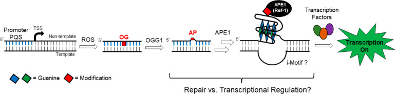
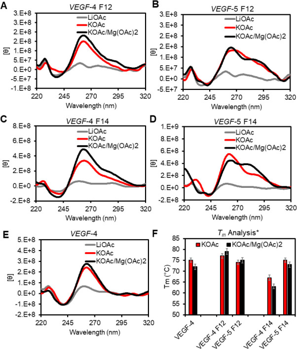
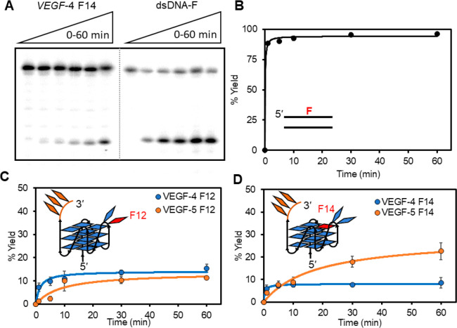
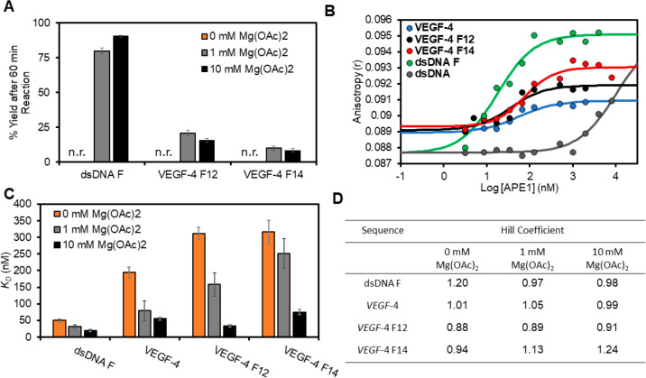
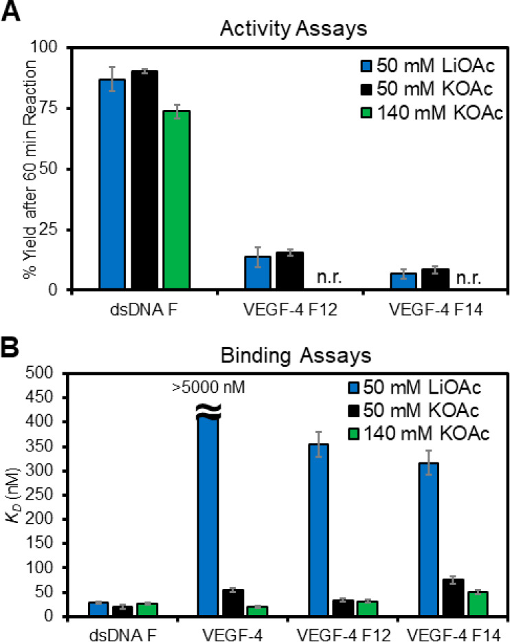
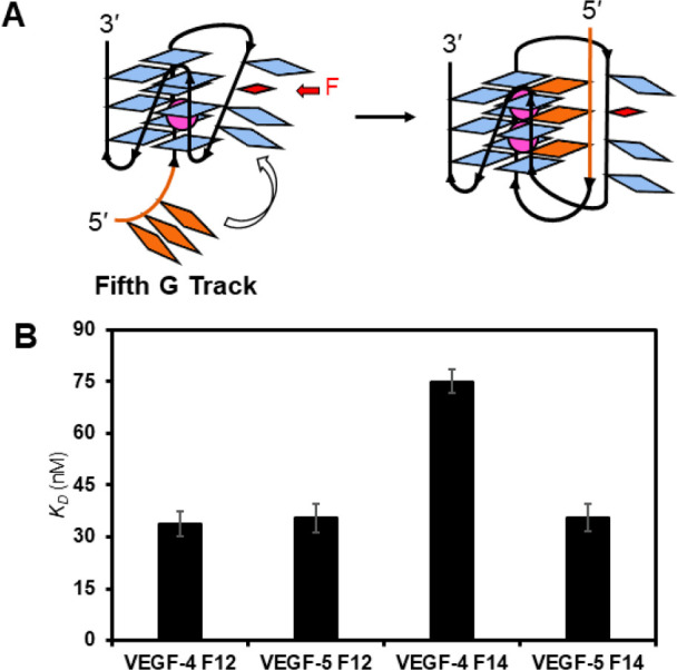
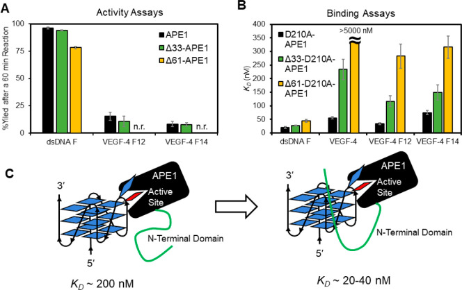
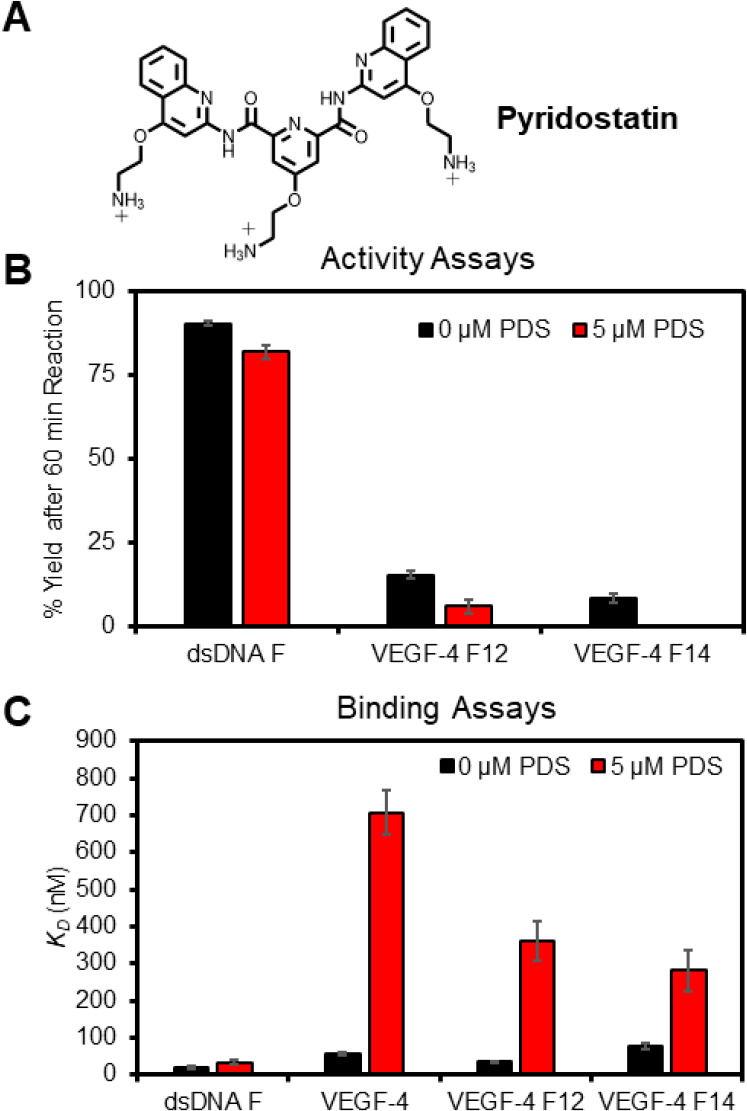
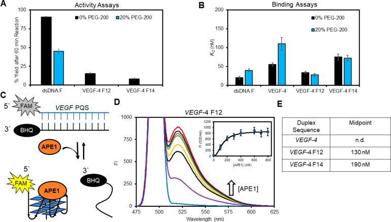
The base excision repair enzyme apurinic/apyrimidinic endonuclease-1 (APE1) is also engaged in transcriptional regulation. APE1 can function in both pathways when the protein binds to a promoter G-quadruplex (G4) bearing an abasic site (modeled with tetrahydrofuran, F) that leads to enzymatic stalling on the non-canonical fold to recruit activating transcription factors. Biochemical and biophysical studies to address APE1's binding and catalytic activity with the vascular endothelial growth factor (VEGF) promoter G4 are lacking, and the present work provides insight on this topic. Herein, the native APE1 was used for cleavage assays, and the catalytically inactive mutant D210A was used for binding assays with double-stranded DNA (dsDNA) versus the native G4 or the G4 with F at various positions, revealing dependencies of the interaction on the cation concentrations K+ and Mg2+ and the N-terminal domain of the protein. Assays in 0, 1, or 10 mM Mg2+ found that dsDNA and G4 substrates required the cation for both binding and catalysis, in which G4 binding increased with [Mg2+]. Studies with 50 versus physiological 140 mM K+ ions showed that F-containing dsDNA was bound and cleaved by APE1; whereas, the G4s with F were poorly cleaved in low salt and not cleaved at all at higher salt while the binding remained robust. Using Δ33 or Δ61 N-terminal truncated APE1 proteins, the binding and cleavage of dsDNA with F was minimally impacted; in contrast, the G4s required the N-terminus for binding and catalysis is nearly abolished without the N-terminus. With this knowledge, we found APE1 could remodel the F-containing VEGF promoter dsDNA→G4 folds in solution. Lastly, the addition of the G4 ligand pyridostatin inhibited APE1 binding and cleavage of F-containing G4s but not dsDNA. The biological and medicinal chemistry implications of the results are discussed.

Keywords: APE1; DNA remodeling; G-Quadruplex; binding assays; gene regulation; pyridostatin.


Conflict of interest statement

Conflict of Interest. No conflicts of interest are declared in this work.

Figures

Figure 1
Figure 1
Multifunctional nuclease APE1 is a member of the base excision repair pathway and can function in transcriptional regulation. The present work asks what molecular properties of the DNA and protein lead to the crossover from one function to the other.
Figure 2
Figure 2
Analysis of the impact of K+ and Mg2+ cations on the folding and stability of the VEGF G4s. (A–E) Inspection of G4 folding by CD spectroscopy in different buffer systems at 22 °C. (F) A plot of the Tm values measured and the respective errors from three independent analyses in the different buffer systems. The buffer and salt compositions represented by the labels are as follows: LiOAc = 20 mM Tris pH 7.4 at 22 °C with 50 mM LiOAc. KOAc = 20 mM Tris pH 7.4 at 22 °C with 50 mM KOAc; KOAc/Mg(OAc)2 = 20 mM Tris pH 7.4 at 22 °C 50 mM KOAc and 10 mM Mg(OAc)2. *The Tm values were recorded in 20 mM KPi buffer at pH 7.4.
Figure 3
Figure 3
Monitoring the APE1-catalyzed cleavage of an AP site in the VEGF sequence by PAGE analysis to identify position- and context-dependent yields. (A) Example time-dependent PAGE analysis of APE1-catalyzed strand scission at an F site in the dsDNA or G4 contexts. The yield vs time profiles for cleavage in the (B) dsDNA context with F at positions 12 (dsDNA F), as well as four- and five-track G4 contexts with F at position (C) 12 or (D) 14. The reactions were conducted by preincubating 10 nM DNA in 20 mM Tris pH 7.5 (37 °C) with 50 mM KOAc, 10 mM Mg(OAc)2, and 1 mM DTT present at 37 °C for 30 min. After the preincubation, APE1 was added to a 3 nM final concentration, and the reactions were allowed to progress to the desired time and then quenched by adding a stop solution and heating at 65 °C for 20 min before PAGE analysis.
Figure 4
Figure 4
Endonuclease activity and fluorescence anisotropy binding assays for the interaction between APE1 and the VEGF PQS dsDNA or G4 folds demonstrate a Mg2+ dependency. (A) The [Mg2+]-dependent activity for native APE1 on the F-containing VEGF substrates was measured after 60 min. (B) Binding plots with log[APE1] vs anisotropy (r) for the analysis without Mg2+ present. (C) KD values were obtained by fitting the sigmoidal binding curves with the appropriate Hill equation. The 0 mM Mg2+ data were obtained from WT APE1, while the 1 and 10 mM Mg2+ data were obtained using the catalytically inactive D210A-APE1 mutant. The errors represent the standard deviation of triplicate trials. (D) Table of Hill coefficients obtained from the fits. The studies were conducted in 20 mM Tris, 50 mM KOAc, and 0, 1, or 10 mM Mg(OAc)2, 1 mM DTT at pH 7.5 for the activity assays at 37 °C and pH 7.4 for the binding assays at 22 °C. The analyses were conducted in triplicate, and the errors represent the standard deviation of the data points. n.r. = no reaction.
Figure 5
Figure 5
Monovalent cation dependency on the APE1 activity and binding of D210A-APE1 with the VEGF PQSs as duplexes or G4s. (A) Endonuclease yields for APE1 cleaving an F in the dsDNA and G4 contexts in 50 mM Li+ (blue), 50 mM K+ (black), or 140 mM K+ buffers (green). (B) Plots of the KD values measured for D210A-APE1 binding the VEGF dsDNA and G4 structural contexts studied. The analyses were conducted in triplicate and the errors represent the standard deviation of the data points. n.r. = no reaction.
Figure 6
Figure 6
Fifth G track in the VEGF G4 can facilitate (A) folding and (B) APE1 binding to F-containing folds. The analyses were conducted in triplicate, and the errors represent the standard deviation of the data points.
Figure 7
Figure 7
N-Terminal disordered domain of APE1 is required for maximal activity and the lowest KD values for the protein interacting with the VEGF G4s. (A) Cleavage yields after 60 min when full-length APE1 and its N-terminal truncated forms Δ33-APE1 or Δ61-APE1 were allowed to react with an F in dsDNA or the VEGF G4 folds. (B) Binding of N-terminal domain truncated and catalytically inactive double mutants (D210A-APE1, Δ33-D210A-APE1, or Δ61-D210A-APE1) with F-containing G4 and dsDNA substrates. (C) Model illustrating the N-terminal domain enables stronger interactions between the APE1 protein and G4 DNA. All analyses were conducted in 20 mM Tris, 50 mM KOAc, 10 mM Mg(OAc)2, and 1 mM DTT buffers at pH 7.5 and 37 °C for the activity assays and pH 7.4 and 22 °C for the binding assays. The analyses were conducted in triplicate, and the errors represent the standard deviation of the data points. n.r. = no reaction.
Figure 8
Figure 8
G4 ligand PDS impacts APE1 endonuclease activity and D210A-APE1 binding of VEGF G4 DNA strands. (A) Structure of PDS. (B) Cleavage yields were measured after a 60 min reaction. (C) Binding constants for the VEGF sequences studied. The analyses were conducted in triplicate, and the errors represent the standard deviation of the data points. n.r. = no reaction.
Figure 9
Figure 9
Presence of 20% PEG-200 alters the APE1 VEGF G4 interactions to favorably allow the protein to remodel the DNA from dsDNA to G4 DNA. (A) APE1 activity yields determined 60 min post-incubation with and without PEG-200 present. (B) Binding of D210A-APE1 to the VEGF substrates with and without PEG-200 present. (C) Schematic of the FRET assay to monitor D210A-APE1 remodeling of dsDNA to G4 DNA. (D) Example FAM fluorescence spectra recorded as D210A-APE1 was titrated into a solution of VEGF-4 F12 dsDNA; the inset proves the fluorescence intensity at 520 nm that was fit to determine the binding midpoint. (E) Binding midpoints were obtained from the FRET assay for the VEGF sequences studied.

Similar articles

Cited by

References

    1. Fleming A. M.; Burrows C. J. Interplay of guanine oxidation and G-quadruplex folding in gene promoters. J. Am. Chem. Soc. 2020, 142, 1115–1136. 10.1021/jacs.9b11050. - DOI - PMC - PubMed
    1. Wang R.; Hao W.; Pan L.; Boldogh I.; Ba X. The roles of base excision repair enzyme OGG1 in gene expression. Cell. Mol. Life Sci. 2018, 75, 3741–3750. 10.1007/s00018-018-2887-8. - DOI - PMC - PubMed
    1. Fong Y. W.; Cattoglio C.; Tjian R. The intertwined roles of transcription and repair proteins. Mol. Cell 2013, 52, 291–302. 10.1016/j.molcel.2013.10.018. - DOI - PMC - PubMed
    1. Antoniali G.; Malfatti M. C.; Tell G. Unveiling the non-repair face of the base excision repair pathway in RNA processing: A missing link between DNA repair and gene expression?. DNA Repair 2017, 56, 65–74. 10.1016/j.dnarep.2017.06.008. - DOI - PubMed
    1. Fleming A. M.; Burrows C. J. 8-Oxo-7,8-dihydroguanine, friend and foe: Epigenetic-like regulator versus initiator of mutagenesis. DNA Repair 2017, 56, 75–83. 10.1016/j.dnarep.2017.06.009. - DOI - PMC - PubMed