. 2021 Dec 20;4(12):8248-8258.
doi: 10.1021/acsabm.1c00794. Epub 2021 Nov 17.

Preventing Pseudomonas aeruginosa Biofilms on Indwelling Catheters by Surface-Bound Enzymes

Affiliations

Preventing Pseudomonas aeruginosa Biofilms on Indwelling Catheters by Surface-Bound Enzymes

Dalal Asker et al. ACS Appl Bio Mater. .

Abstract

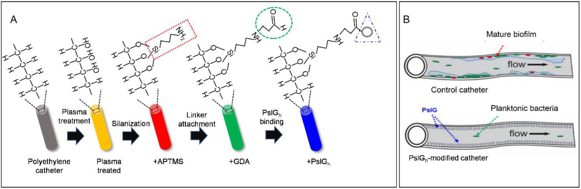
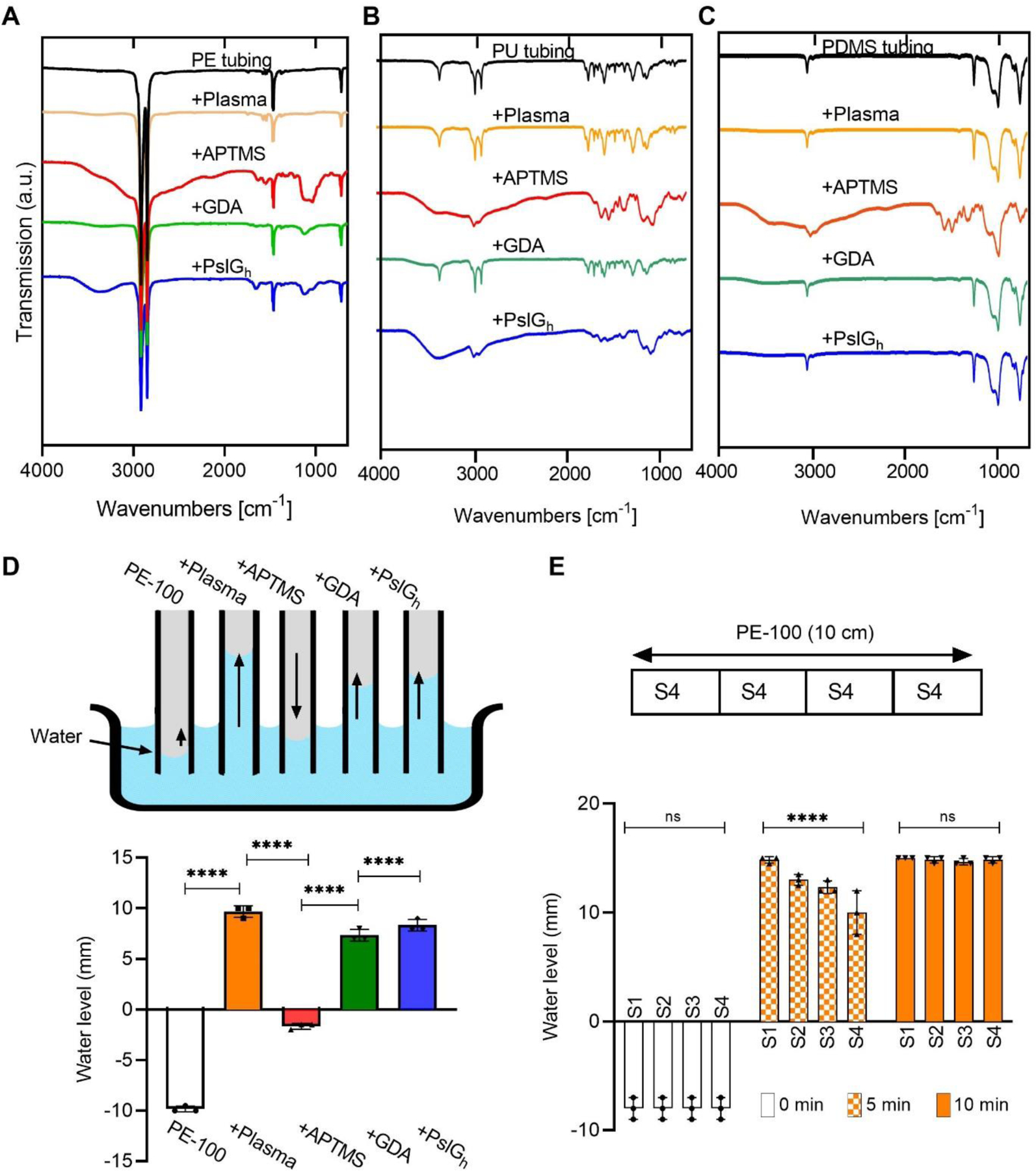
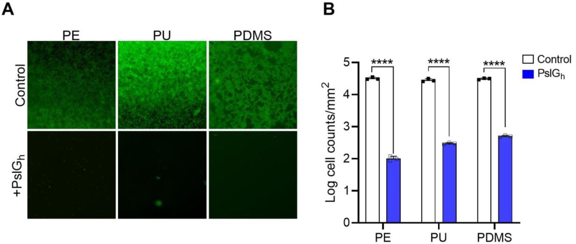
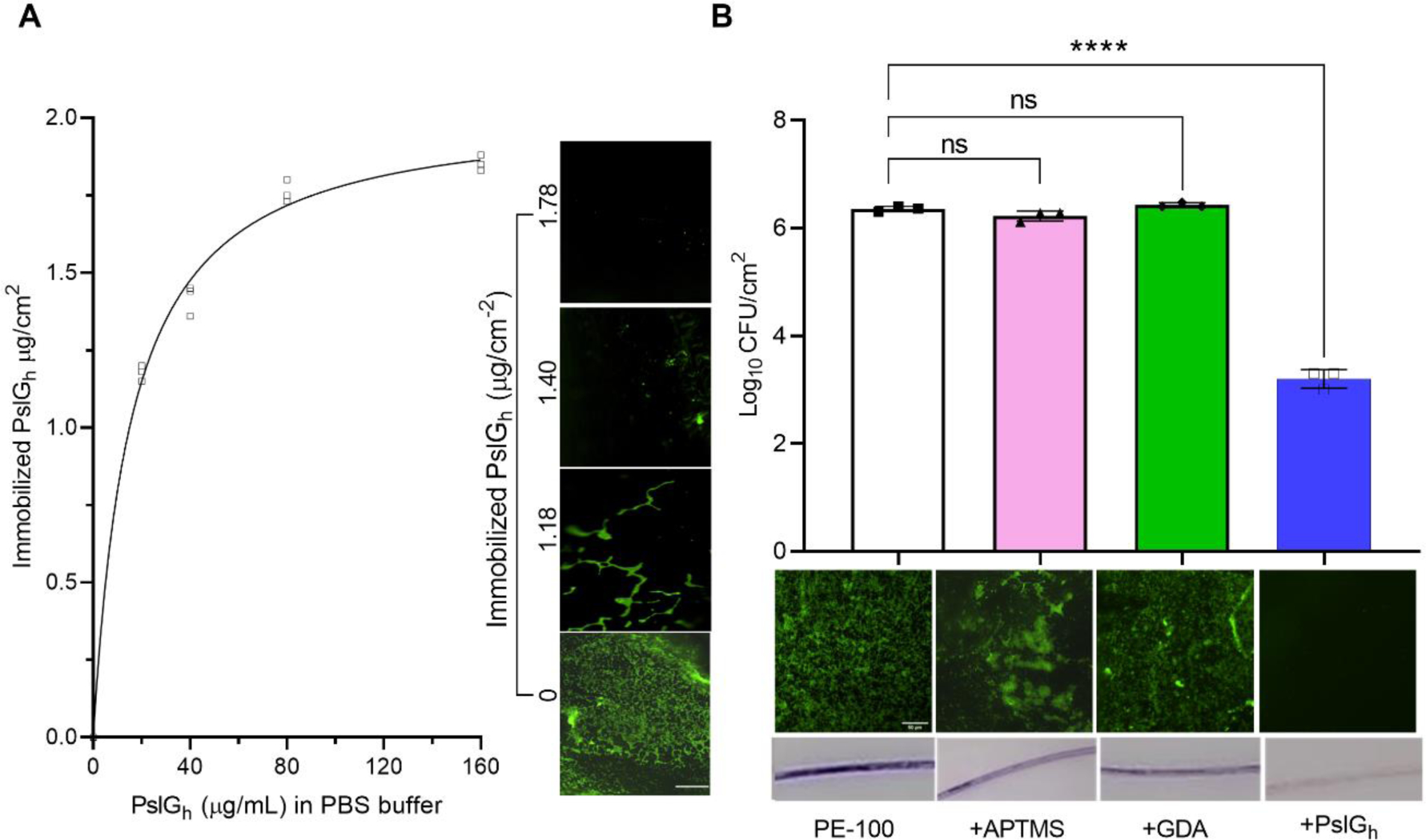
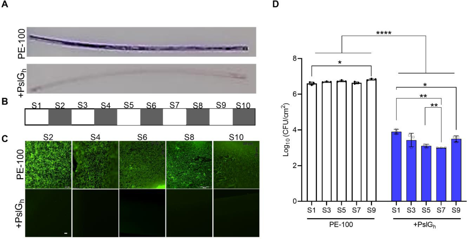
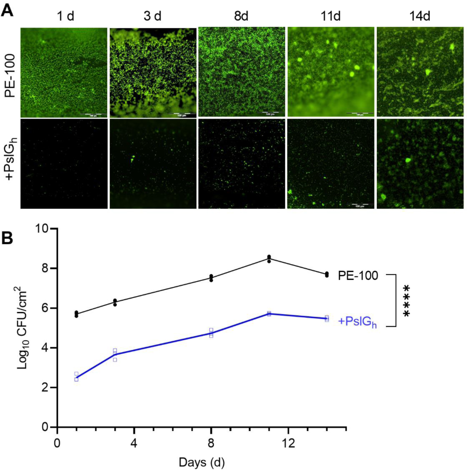
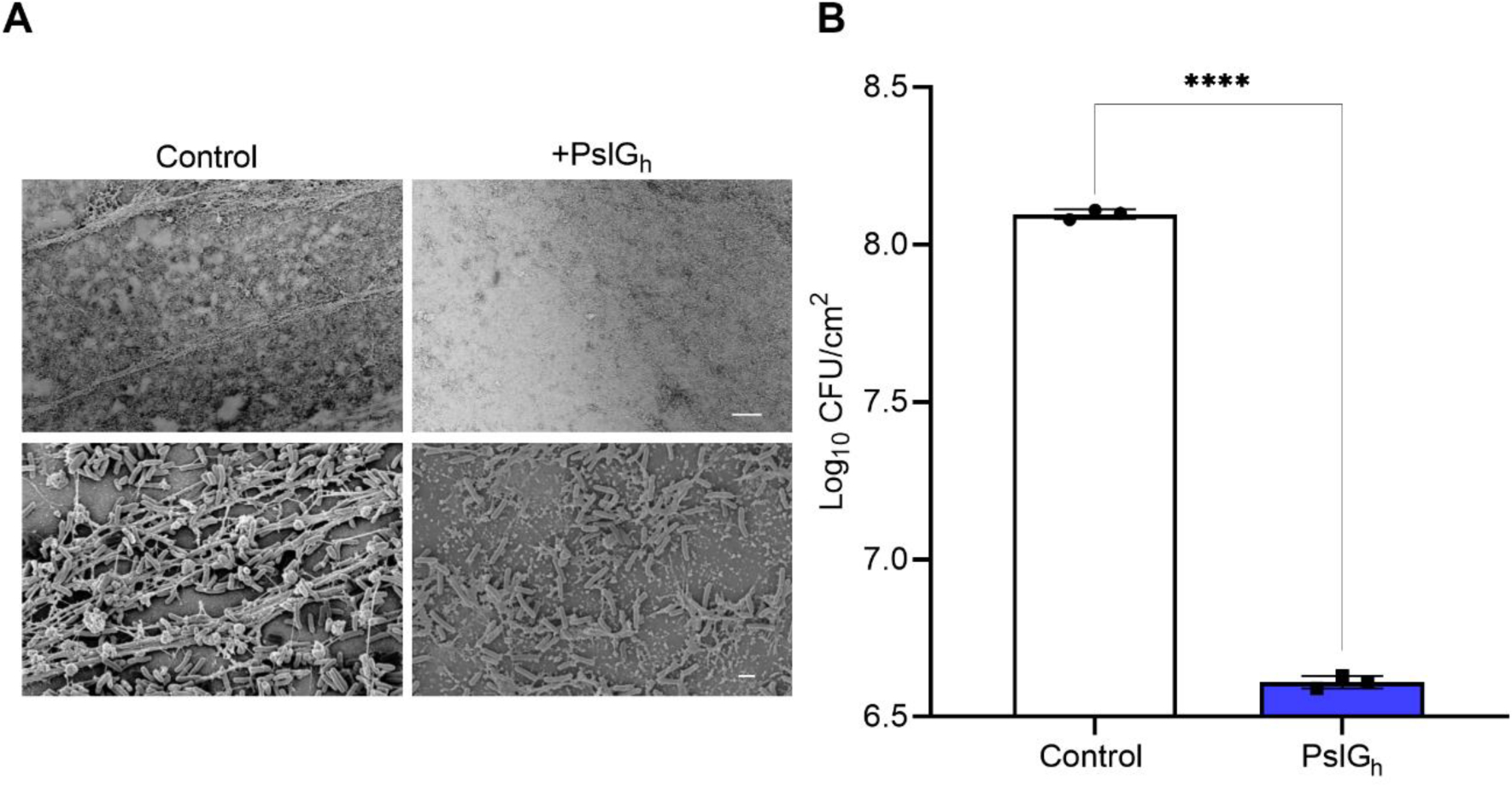
Implanted medical devices such as central venous catheters are highly susceptible to microbial colonization and biofilm formation and are a major risk factor for nosocomial infections. The opportunistic pathogen Pseudomonas aeruginosa uses exopolysaccharides, such as Psl, for both initial surface attachment and biofilm formation. We have previously shown that chemically immobilizing the Psl-specific glycoside hydrolase, PslGh, to a material surface can inhibit P. aeruginosa biofilm formation. Herein, we show that PslGh can be uniformly immobilized on the lumen surface of medical-grade, commercial polyethylene, polyurethane, and polydimethylsiloxane (silicone) catheter tubing. We confirmed that the surface-bound PslGh was uniformly distributed along the catheter length and remained active even after storage for 30 days at 4 °C. P. aeruginosa colonization and biofilm formation under dynamic flow culture conditions in vitro showed a 3-log reduction in the number of bacteria during the first 11 days, and a 2-log reduction by day 14 for PslGh-modified PE-100 catheters, compared to untreated catheter controls. In an in vivo rat infection model, PslGh-modified PE-100 catheters showed a ∼1.5-log reduction in the colonization of the clinical P. aeruginosa ATCC 27853 strain after 24 h. These results demonstrate the robust ability of surface-bound glycoside hydrolase enzymes to inhibit biofilm formation and their potential to reduce rates of device-associated infections.

Keywords: Pseudomonas aeruginosa; PslGh; bacterial biofilms; biomaterials; catheters; enzyme immobilization; glycoside hydrolases; medical device infection.


Figures

Figure 1.
Figure 1.
(A) Schematic illustration of the sequence of steps involved in enzyme immobilization: surface activation by plasma treatment; amine (NH2) functionalization with 3-aminopropyltrimethoxysilane (APTMS) via silanization; activation of the catheter lumen surface using the bifunctional agent (GDA) under the formation of Schiff base and finally coupling of the enzyme. (B) Chemical immobilization of PslGh prevents P. aeruginosa biofilm formation on the luminal surface of catheter tubing. Schematic representation demonstrating cell attachment and biofilm formation on untreated catheters (top) and inhibition of biofilm formation when the catheter is treated with PslGh (bottom).
Figure 2.
Figure 2.
Chemical and physical characterization of the lumen of commercial polymer tubing and a medical grade catheter. (A-C) ATR-FTIR spectra showing the surface chemical groups lining the PE, PU and PDMS tubing lumen during immobilization; (D) Variation in the relative water level inside PE tubing after each immobilization step. A rise in the water level within catheter lumen is indicative of hydrophilic character; and (E) Relative displacement of water inside 4 lumen segments (S1-S4) of a 10cm long PE-100 catheter after treatment with atmospheric plasma for 10 min. ****P ≤ 0.0001. NS, no significant difference.
Figure 3.
Figure 3.
Bound PslGh prevents P. aeruginosa biofilm formation on the luminal surface of polymer tubing. (A) Florescence images showing the inhibition of biofilm formation by bound PslGh on the luminal surface of PE, PU and PDMS tubing, but not for the untreated control. (B) Corresponding cell counts (per cm2) calculated from the image analysis of surfaces in (A). ****P ≤ 0.0001.
Figure 4.
Figure 4.
PslGh-modified PE-100 catheter inhibits biofilm formation (A) Effective concentration of immobilized PslGh required for biofilm inhibition. Effect of PslGh concentration (μg/mL) in immobilization solution on its binding capacity to catheter luminal surface (μg/cm) (left). Effect of immobilized PslGh surface density on biofilm formation of Pseudomonas aeruginosa PAO1 (right). (B) Anti-biofilm activity of the PE catheter lumen with bound PslGh, relative to the untreated control, or APTMS and GDA functionalized surfaces. (A) Colony forming units per cm2 (CFU) (top), corresponding florescence images (middle), and crystal violet (CV) stained images (bottom). All measurements were acquired after incubation for 24 h in bacterial culture. Biofilms were stained with SYTOX Green (middle) and CV (bottom) (scale bar, 50 μm). ****P ≤ 0.001. NS, no significant difference.
Figure 5.
Figure 5.
Antibiofilm activity of the lumen of high density PslGh-bound 10 cm PE-100 catheters against P. aeruginosa. (A) Photos of CV stained 10 cm PE-100 catheters; (B) Fluorescence images of 5 alternating segments (i.e., S2, 4, 6, 8 and 10); and (C) Colony forming units per cm2 (CFU) of the other 5 alternating segments (i.e., S1, 3, 5, 7 and 9). All measurements were acquired after incubation for 24 h in bacterial culture. Biofilms were stained with CV in (A) and with SYTOX Green in (B) (scale bar, 50 μm). *P ≤ 0.05, **P ≤ 0.01, ****P ≤ 0.0001.
Figure 6.
Figure 6.
Inhibition of P. aeruginosa biofilm formation by PslGh bound to the lumen surface of PE-100 catheter under dynamic flow culture conditions. (A) Fluorescence images of biofilm growth for up to 14 days on the lumen surface of untreated (top) and PslGh-bound PE-100 catheter (bottom). (B) Colony forming units (CFU/cm2) of P. aeruginosa PAO1 on the lumen surface of untreated and PslGh-bound PE-100 catheter after 14 d incubation. Biofilms were stained with SYTOX Green in (A) (scale bar, 100 μm). ****P ≤ 0.0001.
Figure 7.
Figure 7.
In vivo inhibition of biofilm formation of P. aeruginosa ATCC 27853 on the PslGh-treated lumen surface of PE catheter. (A) SEM images of biofilm growth on untreated and treated PE-100 catheters at low magnification (1,000X), and (B) corresponding bacterial burden showing ~ 2 log CFU /cm2 reduction in P. aeruginosa ATCC 27853 cells after 1d of placement in vivo. The scale bars represent 20 μm, respectively. ****P ≤ 0.0001.

References

    1. Costerton J; Stewart PS; Greenberg E, Bacterial biofilms: a common cause of persistent infections. Science 1999, 284 (5418), 1318–1322. - PubMed
    1. Del Pozo J; Patel R, The challenge of treating biofilm-associated bacterial infections. Clinical Pharmacology & Therapeutics 2007, 82 (2), 204–209. - PubMed
    1. Percival SL; Suleman L; Vuotto C; Donelli G, Healthcare-associated infections, medical devices and biofilms: risk, tolerance and control. Journal of medical microbiology 2015, 64 (4), 323–334. - PubMed
    1. Wolcott R; Rhoads D; Bennett M; Wolcott B; Gogokhia L; Costerton J; Dowd S, Chronic wounds and the medical biofilm paradigm. Journal of wound care 2010, 19 (2), 45–53. - PubMed
    1. Flemming H-C; Wingender J, The biofilm matrix. Nature Reviews Microbiology 2010, 8 (9), 623. - PubMed

Publication types

Substances

LinkOut - more resources