Review
. 2021 Jun 10;2(1):17.
doi: 10.1186/s43556-021-00036-1.

G-protein coupled receptor, PI3K and Rho signaling pathways regulate the cascades of Tau and amyloid-β in Alzheimer's disease

Affiliations
Review

G-protein coupled receptor, PI3K and Rho signaling pathways regulate the cascades of Tau and amyloid-β in Alzheimer's disease

Smita Eknath Desale et al. Mol Biomed. .

Abstract

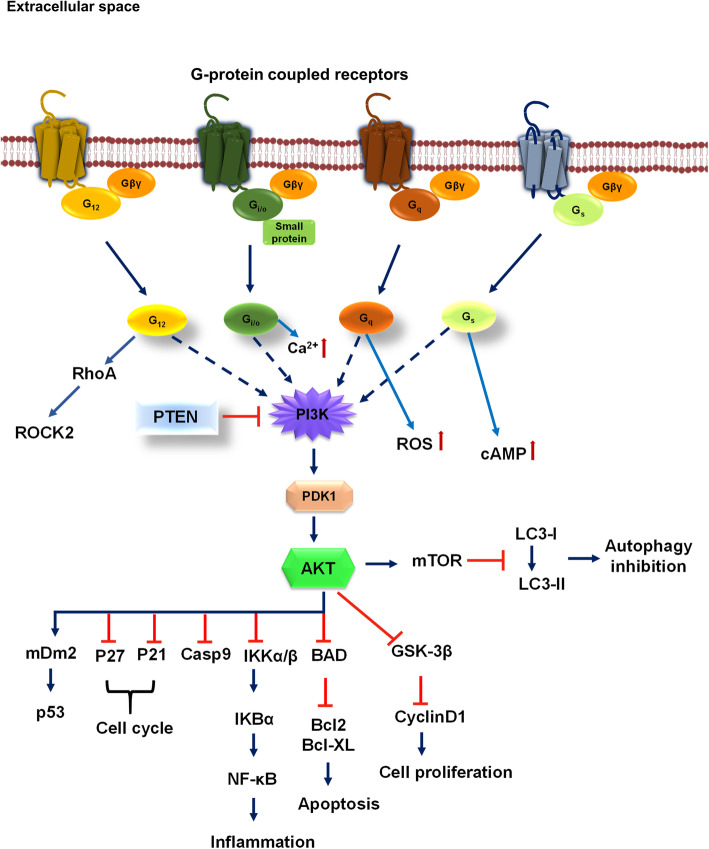
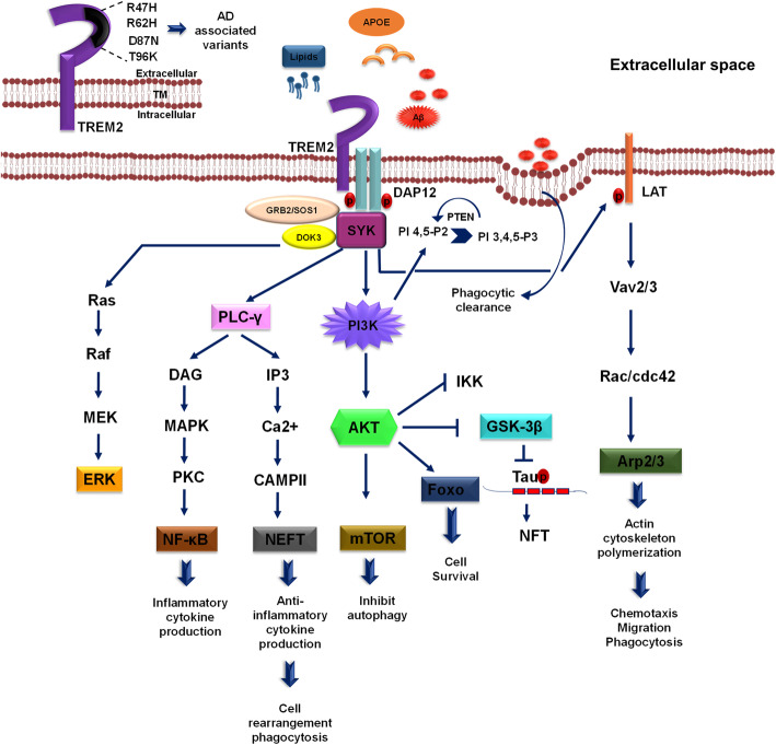
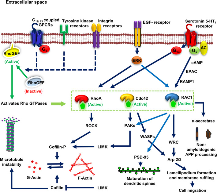
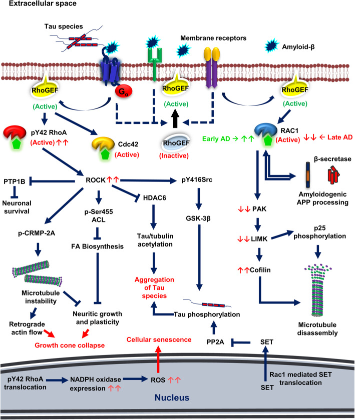
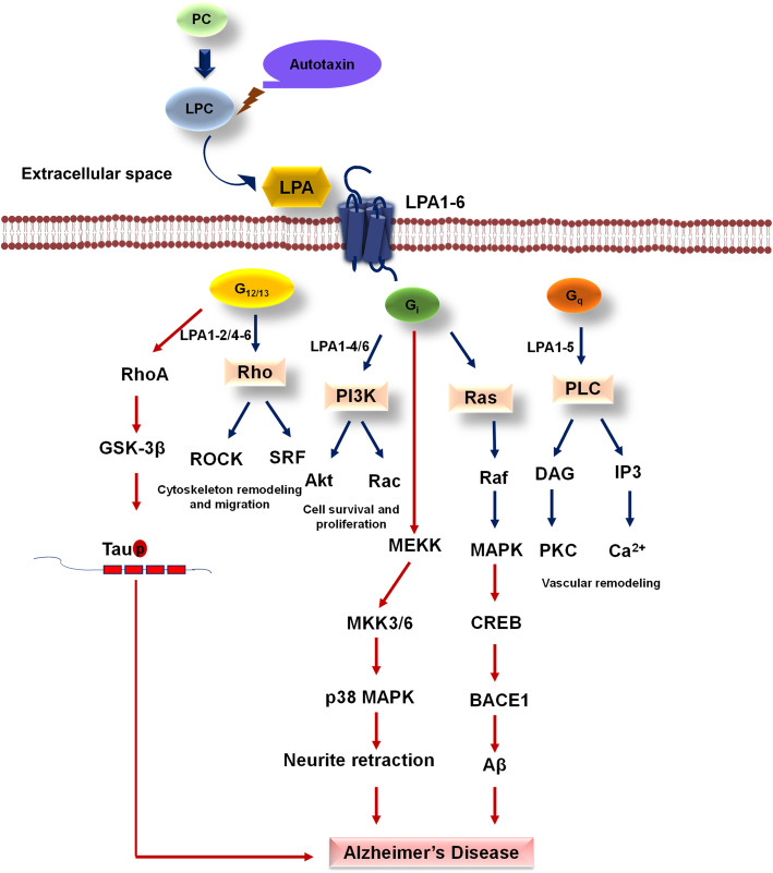
Alzheimer's disease is a progressive neurodegenerative disease characterized by the presence of amyloid-β plaques in the extracellular environment and aggregates of Tau protein that forms neurofibrillary tangles (NFTs) in neuronal cells. Along with these pathological proteins, the disease shows neuroinflammation, neuronal death, impairment in the immune function of microglia and synaptic loss, which are mediated by several important signaling pathways. The PI3K/Akt-mediated survival-signaling pathway is activated by many receptors such as G-protein coupled receptors (GPCRs), triggering receptor expressed on myeloid cells 2 (TREM2), and lysophosphatidic acid (LPA) receptor. The signaling pathway not only increases the survival of neurons but also regulates inflammation, phagocytosis, cellular protection, Tau phosphorylation and Aβ secretion as well. In this review, we focused on receptors, which activate PI3K/Akt pathway and its potential to treat Alzheimer's disease. Among several membrane receptors, GPCRs are the major drug targets for therapy, and GPCR signaling pathways are altered during Alzheimer's disease. Several GPCRs are involved in the pathogenic progression, phosphorylation of Tau protein by activation of various cellular kinases and are involved in the amyloidogenic pathway of amyloid-β synthesis. Apart from various GPCR signaling pathways, GPCR regulating/ interacting proteins are involved in the pathogenesis of Alzheimer's disease. These include several small GTPases, Ras homolog enriched in brain, GPCR associated sorting proteins, β-arrestins, etc., that play a critical role in disease progression and has been elaborated in this review.

Keywords: Alzheimer’s disease; GPCR; LPA; PI3K/Akt; Rho GTPase; TREM2; Tau.


Conflict of interest statement

The authors declare that they have no competing interests.

Figures

Fig. 1
Fig. 1
GPCR influence PI3K/Akt signaling. Among various GPCR G12/13, Gi/o, Gq, Gs are known to activate PI3K/Akt cell survival pathway via its Gα or Gβγ subunit. Along with PI3K/Akt signaling G12/13 activate RhoA/ROCK pathway, Gi/o involves in maintaining intracellular levels of Ca2+, Gq with the ROS production, and Gas with secondary messenger cAMP. The regulation of PI3K is carried out by PTEN, a phosphatase of PI 3, 4, 5-P3, which inhibits PI3K directly. Upon activation of PI3K, Akt phosphorylation occurs via PDK1 kinase, followed by mTOR pathway activation, which inhibits autophagy response by inhibiting the conversion of LC3-l to LC3-ll. Akt specifically affects various downstream signaling pathway such as inhibition of GSK-3β, BAD, IKK, Casp9, P27, P21, and activates mDm2, which is involved in the p53-mediated response. This indicates a wide importance of Akt signaling in various cellular pathways
Fig. 2
Fig. 2
PI3K/Akt signaling-mediated by TREM2. DAP12-mediated TREM2 signaling have been discussed in the figure. The possible ligands with respect to Alzheimer’s disease for TREM2 are lipids, APOE as well as amyloid-β. In the physiological condition, upon tyrosine phosphorylation of DAP12 ITAM leads to recruitment of Syk kinase along with the GRB/SOS1 and DOK3, which activates downstream signaling pathways of ERK, PLC-γ, PI3K/Akt as well as Vav. The activation of PLC-γ induces inflammatory cytokine response via DAG stimulation and anti-inflammatory cytokine response along with cell rearrangement and phagocytosis via elevating Ca2+ response in the cell through IP3 activation. The main survival pathway of cell PI3K/Akt primarily activates mTOR pathway and Foxo that inhibit autophagy and induce cell survival, inhibits GSK-3β that eventually reduce Tau phosphorylation in Alzheimer’s disease and suppress IKK, which is involved in the inflammatory response. PI3K activation increase formation of PI 3,4,5-P3, an important phosphoinositide in the process of phagocytosis and migration. Syk recruitment activates Vav2/3 molecule via LAT phosphorylation, which induces actin cytoskeleton polymerization important for chemotaxis, migration, and phagocytosis. TREM2 is one of the disease-linked genes in Alzheimer’s disease, the AD-related variant generally present in Ig-like extracellular domain of the receptor, which includes R47H, R62H, D87N, T96K
Fig. 3
Fig. 3
Physiological activation and signaling of Rho-GTPases. Several membrane receptors, such as G12/13 coupled GPCRs, Tyrosine kinase receptors, Integrin receptors, etc., are involved in the activation of Rho-GEFs. The active form of Rho-GEF binds to the membrane and are involved in activation of Rho-GTPases (mostly RhoA, Rac1 and Cdc42) through GTP catalysis. Epidermal growth factor receptor is involved in ERK-mediated phosphorylation of RhoA and Rac1 and its activation. Serotonin 5-HT4 receptor activates cAMP, EPAC and RAMP1 via Gs signaling and activates Rac1 signaling that is involved in the promotion of non-amyloidogenic APP processing, dendritic spine formation (PSD-95 expression) in mature neurons, lamellipodium formation and membrane ruffling for migration. Serotonin 5-HT4 receptor also activates RhoA via G13 proteins. Activated RHO-GTPases are involved in several vital functions such as cell migration, maturation of dendritic spines, neuritic outgrowth, learning, memory, etc., RhoA GTPase activates ROCK kinase that is involved in the phosphorylation of cofilin. Cofilin is involved in the formation of globular actin from filamentous actin (actin depolymerization), and this process is inhibited and reversed upon cofilin phosphorylation. Cofilin also binds tubulin and promotes microtubule instability. Mixed lineage kinases (MLK) and p-21 activated kinases (PAK) are the common effectors of Cdc42 and Rac1. PAKs are involved in the activation of LIM kinase, which also involved in the phosphorylation of cofilin. Wiskott–Aldrich syndrome protein (WASP) and WAVE regulatory complex (WRC), the downstream effectors of Cdc42 and Rac1 respectively activate Arp 2/3 complex which promotes membrane ruffling during migration
Fig. 4
Fig. 4
Pathological signaling of Rho GTPases in Alzheimer’s disease. In Alzheimer’s disease, the extracellular space is filled with senile plaques of amyloid-β and aggregated Tau species. These disordered proteins interact with several surface receptors, which on activation lead to pathological signaling of Rho-GTPases. Rho-GTPases are highly activated (phosphorylated on Y42) on amyloid-β exposure. Hyperactivation of Rho-GTPases leads to increased activation of ROCK kinases. ROCK kinase phosphorylates Src at Tyrosine 416 and activates to activate GSK-3β, an enzyme involved in Tau hyperphosphorylation. ROCK also inhibits HDAC6 activity which ultimately promotes Tau and tubulin acetylation. All these post-translational modifications of Tau lead to aggregation of Tau species. ROCK phosphorylates and inactivates CRMP-2A and ATP citrate lyase (p-Ser 455) (ACL). CRMP-2A are microtubule-associated proteins, and its inactivation led to microtubule instability and retrograde flow of actin filaments. Fatty acid biosynthesis is inhibited by ATP-citrate lyase phosphorylation which ultimately inhibits neurite growth, plasticity and leads to growth cone collapse in neuronal cells. Activated ROCK kinases also inhibit protein tyrosine phosphatase 1B and neuronal survival. Phosphorylated RhoA translocates to the nucleus and promotes the expression of NADPH oxidase, increases ROS levels and cellular senescence. Rac1 GTPase expression level varies at different stages and promotes amyloidogenic APP processing during the later stages of Alzheimer’s disease leading to more amyloid-β secretion and vice versa. At later stages, Rac1 expression level is reduced, and subsequent effectors PAK and LIMK activities are also downregulated. This condition leads to the imbalance in cofilin (active) and its phosphorylated form (inactive). Excessive cofilin activity causes actin depolymerization and microtubule instability. Under AD conditions, Rac1 also promotes translocation of SET proteins from the nucleus to the cytoplasm that acts as phosphatase inhibitors and ultimately promotes phosphorylation and aggregation of Tau species
Fig. 5
Fig. 5
GPCR in Lysophosphatidic acid cycle. LPA production through LPC via autotaxin enzyme exerts its function through cell surface GPCR mainly G12/13, Gi/o, and Gαq protein-mediated signals. Amongst six different LPA, LPA1-2/4-6 exert their function through Gα12/13 and follows Rho-mediated cytoskeleton remodeling which assists cellular migration. LPA1-4/6 exert their function through Gi/o, which is responsible for activating PI3K and Ras-mediated MAPK signaling for cell survival and proliferation. Gαq protein-mediated signal activate the PLC pathway results in vascular remodeling on stimulation by LPA1-5. Defect in signaling through Gα12/13 in AD abnormally increases active GSK-3β, which increases phospho-Tau. Gi leads to dysregulation of BACE1, which impose abnormal Aβ and neurite retraction via p38 MAPK kinases

Similar articles

Cited by

References

    1. DeTure MA, Dickson DW. The neuropathological diagnosis of Alzheimer’s disease. Mol Neurodegener. 2019;14(1):1–18. doi: 10.1186/s13024-019-0333-5. - DOI - PMC - PubMed
    1. Cvetković-Dožić D, Skender-Gazibara M, Dožić S. Neuropathological hallmarks of Alzheimer's disease. Arch Oncol. 2001;9(3):195–199.
    1. Serrano-Pozo A, Frosch MP, Masliah E, Hyman BT. Neuropathological alterations in Alzheimer disease. Cold Spring Harb Perspect Med. 2011;1(1):a006189. doi: 10.1101/cshperspect.a006189. - DOI - PMC - PubMed
    1. Binder LI, Frankfurter A, Rebhun LI. The distribution of tau in the mammalian central nervous system. J Cell Biol. 1985;101(4):1371–1378. doi: 10.1083/jcb.101.4.1371. - DOI - PMC - PubMed
    1. Conde C, Cáceres A. Microtubule assembly, organization and dynamics in axons and dendrites. Nat Rev Neurosci. 2009;10(5):319–332. doi: 10.1038/nrn2631. - DOI - PubMed

LinkOut - more resources