. 2020 Dec 30;1(1):19.
doi: 10.1186/s43556-020-00023-y.

RAD50 deficiency is a predictor of platinum sensitivity in sporadic epithelial ovarian cancers

Affiliations

RAD50 deficiency is a predictor of platinum sensitivity in sporadic epithelial ovarian cancers

Adel Alblihy et al. Mol Biomed. .

Abstract

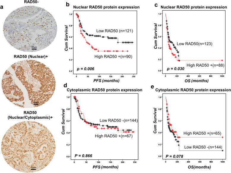
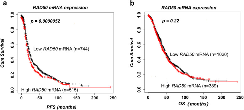
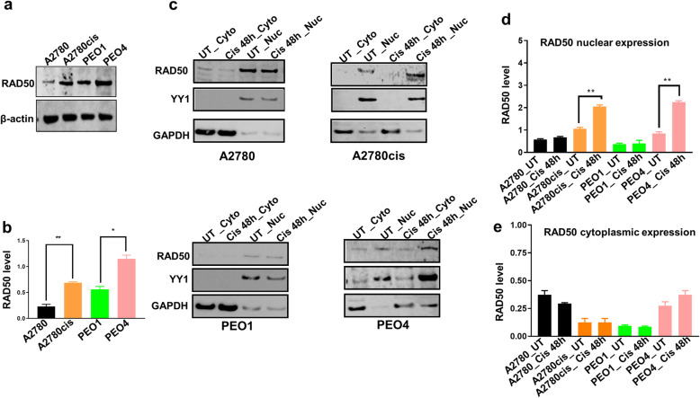
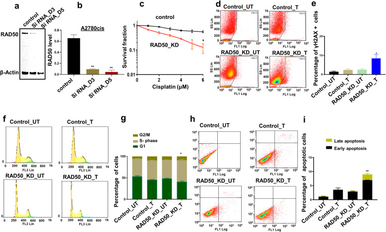
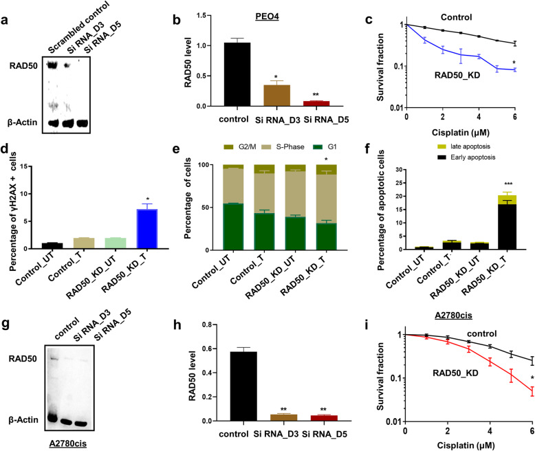
Intrinsic or acquired resistance seriously limits the use of platinating agents in advanced epithelial ovarian cancers. Increased DNA repair capacity is a key route to platinum resistance. RAD50 is a critical component of the MRN complex, a 'first responder' to DNA damage and essential for the repair of DSBs and stalled replication forks. We hypothesised a role for RAD50 in ovarian cancer pathogenesis and therapeutics. Clinicopathological significance of RAD50 expression was evaluated in clinical cohorts of ovarian cancer at the protein level (n = 331) and at the transcriptomic level (n = 1259). Sub-cellular localization of RAD50 at baseline and following cisplatin therapy was tested in platinum resistant (A2780cis, PEO4) and sensitive (A2780, PEO1) ovarian cancer cells. RAD50 was depleted and cisplatin sensitivity was investigated in A2780cis and PEO4 cells. RAD50 deficiency was associated with better progression free survival (PFS) at the protein (p = 0.006) and transcriptomic level (p < 0.001). Basal level of RAD50 was higher in platinum resistant cells. Following cisplatin treatment, increased nuclear localization of RAD50 was evident in A2780cis and PEO4 compared to A2780 and PEO1 cells. RAD50 depletion using siRNAs in A2780cis and PEO4 cells increased cisplatin cytotoxicity, which was associated with accumulation of DSBs, S-phase cell cycle arrest and increased apoptosis. We provide evidence that RAD50 deficiency is a predictor of platinum sensitivity. RAD50 expression-based stratification and personalization could be viable clinical strategy in ovarian cancers.

Keywords: DNA repair; Ovarian cancer; Platinum therapy; Predictive biomarker; RAD50.


Conflict of interest statement

The authors disclose no potential conflicts of interest.

Figures

Fig. 1
Fig. 1
RAD50 protein expression and survival in ovarian cancers. a Immunohistochemical expression of RAD50 in ovarian cancers [‘-‘= no tumour expression, ‘+’ = tumour expression]. b Kaplan-Meier curve for RAD50 nuclear protein expression and progression free survival (PFS) in ovarian cancer. c RAD50 nuclear protein expression and overall survival (OS) in ovarian cancer. d Kaplan-Meier curve for RAD50 cytoplasmic protein expression and PFS in ovarian cancer. e RAD50 cytoplasmic protein expression and OS in ovarian cancer
Fig. 2
Fig. 2
RAD50 mRNA expression and survival in ovarian cancers. a The association between RAD50 mRNA expression and PFS. b The association between RAD50 mRNA expression and OS
Fig. 3
Fig. 3
RAD50 protein expression in ovarian cancer cells. a Basal RAD50 protein level in A2780, A2780cis, PEO1 and PEO4 cell lines. b Quantification of RAD50 baseline levels in A2780, A2780cis, PEO1 and PEO4 cell lines. c Western blot of RAD50 protein level in nuclear (Nuc) and cytoplasmic extracts (Cyto) of A2780, A2780cis, PEO1 and PEO4 treated with 5 μM cisplatin. Nuclear and cytoplasmic lysates collected 48 h post treatment. d Quantification of RAD50 nuclear sub-cellular localization in A2780, A2780cis, PEO1 and PEO4 cell lines. e Quantification of RAD50 cytoplasmic expression in A2780, A2780cis, PEO1 and PEO4 cell lines. YY1 was used as a loading control to the nuclear fractions and GADPH as a loading control for the cytoplasmic fractions. UN = untreated cells. Cis = cisplatin
Fig. 4
Fig. 4
RAD50 depletion and cisplatin sensitivity in A2780cis cells. a RAD50_KD in A2780cis cells. b Quantification of western blot showing RAD50 depletion in A2780cis cells. c Cisplatin sensitivity in A2780cis control and A2780cis_RAD50_KD cells (clonogenic assay). d Representative photomicrograph of yH2AX flow cytometry assay for DSBs in control and RAD50_KD_A2780cis cells treated with cisplatin compared to scrambled controls. e The percentage of γH2Ax positive cells by flow cytometry in control and RAD50_KD_A2780cis cells treated with cisplatin compared to scrambled controls. f Representative photomicrograph of PI flow cytometry assay for cell cycle progression in control and RAD50_KD_A2780cis cells treated with cisplatin compared to scrambled controls. g Cell cycle analysis by flow cytometry in control and RAD50_KD_A2780cis cells treated with cisplatin compared to scrambled controls. h Representative photomicrograph of Annexin-V flow cytometry assay for apoptotic cells in control and RAD50_KD_A2780cis cells treated with cisplatin compared to scrambled controls. i Annexin V analysis by flow cytometry in control and RAD50_KD_A2780cis cells treated with cisplatin compared to scrambled controls. UN = untreated cells. T = treated cells
Fig. 5
Fig. 5
RAD50 depletion and cisplatin sensitivity in PEO4 cells. a RAD50_KD in PEO4 cells. b Quantification of western blot showing RAD50 depletion in PEO4 cells. c Cisplatin sensitivity in A2780cis control and PEO4_RAD50_KD cells (clonogenic assay). d The percentage of γH2Ax positive cells by flow cytometry in control and RAD50_KD_ PEO4 cells treated with cisplatin compared to scrambled controls. e Cell cycle analysis by flow cytometry in control and RAD50_KD_ PEO4 cells treated with cisplatin compared to scrambled controls. f Annexin V analysis by flow cytometry in control and RAD50_KD_ PEO4 cells treated with cisplatin compared to scrambled controls. UN = untreated cells. T = treated cells. g RAD50_KD in A2780cis cells using second construct. h Quantification of western blot showing RAD50 depletion in A2780cis cells. i Cisplatin sensitivity in A2780cis control and A2780cis_RAD50_KD cells (clonogenic assay) using second construct. UN = untreated cells. T = treated cells

References

    1. Mesquita KA, Alabdullah M, Griffin M, Toss MS, Fatah T, Alblihy A, et al. ERCC1-XPF deficiency is a predictor of olaparib induced synthetic lethality and platinum sensitivity in epithelial ovarian cancers. Gynecol Oncol. 2019;153(2):416–424. doi: 10.1016/j.ygyno.2019.02.014. - DOI - PubMed
    1. Spivak G. Nucleotide excision repair in humans. DNA Repair (Amst) 2015;36:13–18. doi: 10.1016/j.dnarep.2015.09.003. - DOI - PMC - PubMed
    1. Scharer OD. Nucleotide excision repair in eukaryotes. Cold Spring Harb Perspect Biol. 2013;5(10):a012609. doi: 10.1101/cshperspect.a012609. - DOI - PMC - PubMed
    1. Lopez-Martinez D, Liang C-C, Cohn MA. Cellular response to DNA interstrand crosslinks: the Fanconi anemia pathway. Cell Mol Life Sci. 2016;73(16):3097–3114. doi: 10.1007/s00018-016-2218-x. - DOI - PMC - PubMed
    1. Basourakos SP, Li L, Aparicio AM, Corn PG, Kim J, Thompson TC. Combination platinum-based and DNA damage response-targeting cancer therapy: evolution and future directions. Curr Med Chem. 2017;24(15):1586–1606. doi: 10.2174/0929867323666161214114948. - DOI - PMC - PubMed

LinkOut - more resources