Review
. 2021 Nov 5;2(1):34.
doi: 10.1186/s43556-021-00047-y.

Targeting cardiomyocyte proliferation as a key approach of promoting heart repair after injury

Affiliations
Review

Targeting cardiomyocyte proliferation as a key approach of promoting heart repair after injury

Shuainan Li et al. Mol Biomed. .

Abstract

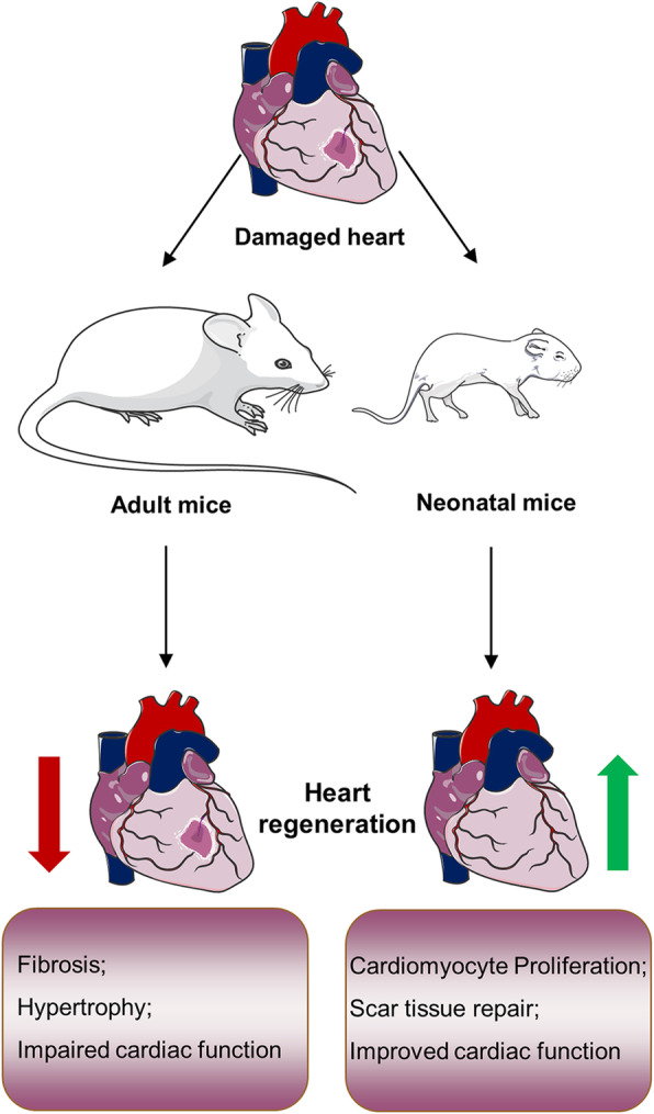
Cardiovascular diseases such as myocardial infarction (MI) is a major contributor to human mortality and morbidity. The mammalian adult heart almost loses its plasticity to appreciably regenerate new cardiomyocytes after injuries, such as MI and heart failure. The neonatal heart exhibits robust proliferative capacity when exposed to varying forms of myocardial damage. The ability of the neonatal heart to repair the injury and prevent pathological left ventricular remodeling leads to preserved or improved cardiac function. Therefore, promoting cardiomyocyte proliferation after injuries to reinitiate the process of cardiomyocyte regeneration, and suppress heart failure and other serious cardiovascular problems have become the primary goal of many researchers. Here, we review recent studies in this field and summarize the factors that act upon the proliferation of cardiomyocytes and cardiac repair after injury and discuss the new possibilities for potential clinical treatment strategies for cardiovascular diseases.

Keywords: Cardiac repair; Cardiomyocyte proliferation; Cardiovascular disease; Heart regeneration; MicroRNAs; Myocardial infarction.


Conflict of interest statement

The authors declare that they have no conflict of interest.

Figures

Fig. 1
Fig. 1
Overview of heart regeneration in mice inside and outside the regeneration window. After cardiac damage, neonatal mice exhibit promotion of cardiomyocyte proliferation, scar tissue repair, and improvement in cardiac function. Conversely, adult mice exhibit myocardial fibrosis, hypertrophy, and impairment of cardiac function
Fig. 2
Fig. 2
A sketch of the working model and interactions of the Hippo, Notch, and NRG1/ErbB signaling pathways. Specifically, Notch signaling pathway activation by Jagged/Delta binding to Notch followed by two protein hydrolysis releases NICD into the nucleus, and then NICD binds to CSL for targeting gene expression. However, Hippo signaling is activated by cascade phosphorylation of core kinases (MST1/MST2, SAV1, LATS1/LATS2, MOB1), which subsequently phosphorylate YAP and TAZ so that they cannot enter the nucleus to bind to TEAD family members. In addition, there is crosstalk between ErbB signaling and Hippo signaling, as ErbB signaling activates ERK and thus phosphorylates the S352 and S274 sites of YAP to promote transcription of target genes; incidentally, this works by acting on the non-classical Hippo/YAP pathway
Fig. 3
Fig. 3
The classical Wnt/β-catenin signaling pathway in cardiac regeneration. After binding of Wnt ligands to their membrane receptors Frizzled and LRP5/6, Dvl inhibits the degradation of the disruption complex and stabilizes β-catenin in the cytoplasm. The increased β-catenin in the nucleus binds to TCF/LEF and regulates the transcription of target genes. In addition, the activation of IGF signaling during this process promotes the stabilization of β-catenin by affecting PI3K/AKT to inhibit the expression of GSK-3β
Fig. 4
Fig. 4
Major factors that target cardiomyocyte proliferation and their possible molecular pathways. Green arrows indicate that cardiomyocyte proliferation or gene expression is promoted, and red solid lines indicate that cardiomyocyte proliferation or gene expression is inhibited. Bcl-2: B-cell lymphoma-2; Btg2: B-cell translocation gene 2; BMP2: bone morphogenetic protein-2; Ccna2: cyclin A2; Ccne1: cyclin E1; Cenpa: centromere protein A; CDK1/2/4: cyclin-dependent kinase 1/2/4; C-Myc: cancer MYC; E2F-1: FoxM1: forkhead box protein M1; pSmad: phosphorylated mothers against DPP homolog; PTEN: phosphatase and tensin homologue deleted on chromosome 10; p27/ p21/ p16: cyclin-dependent kinase inhibitor; SUZ12: suppressor of Zeste 12 homolog; Sirt1: Sirtuin 1; VEGF: vascular endothelial growth factor; Wee1: G2 checkpoint kinase

Similar articles

Cited by

References

    1. Mathers CD, Loncar D. Projections of global mortality and burden of disease from 2002 to 2030. PLoS Med. 2006;3(11):e442. doi: 10.1371/journal.pmed.0030442. - DOI - PMC - PubMed
    1. Pagidipati NJ, Gaziano TA. Estimating deaths from cardiovascular disease: a review of global methodologies of mortality measurement. Circulation. 2013;127(6):749–756. doi: 10.1161/CIRCULATIONAHA.112.128413. - DOI - PMC - PubMed
    1. Ohira T, Iso H. Cardiovascular disease epidemiology in Asia: an overview. Circ J. 2013;77(7):1646–1652. doi: 10.1253/circj.cj-13-0702. - DOI - PubMed
    1. Benjamin EJ, Muntner P, Alonso A, Bittencourt MS, Callaway CW, Carson AP, et al. Heart disease and stroke statistics-2019 update: a report from the American heart association. Circulation. 2019;139(10):e56–e528. doi: 10.1161/CIR.0000000000000659. - DOI - PubMed
    1. Virani SS, Alonso A, Benjamin EJ, Bittencourt MS, Callaway CW, Carson AP, et al. Heart disease and stroke statistics-2020 update: a report from the American heart association. Circulation. 2020;141(9):e139–e596. doi: 10.1161/CIR.0000000000000757. - DOI - PubMed

LinkOut - more resources