

Extracellular vesicles (exosomes and ectosomes) play key roles in the pathology of brain diseases
- PMID: 35006460
- PMCID: PMC8607397
- DOI: 10.1186/s43556-021-00040-5
Extracellular vesicles (exosomes and ectosomes) play key roles in the pathology of brain diseases
Abstract
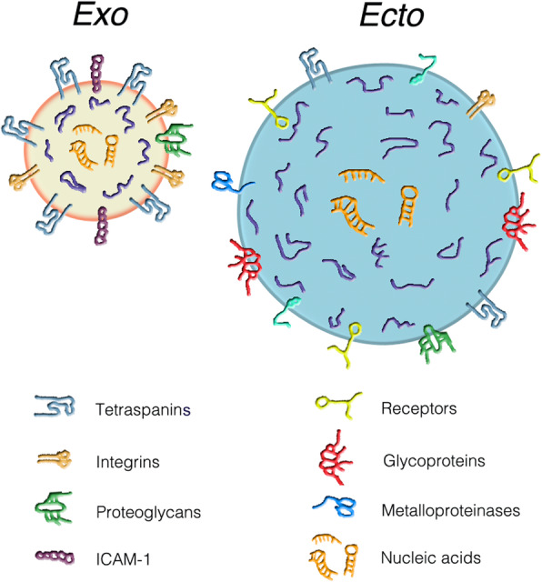
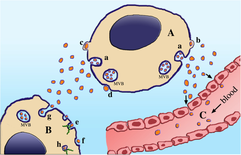
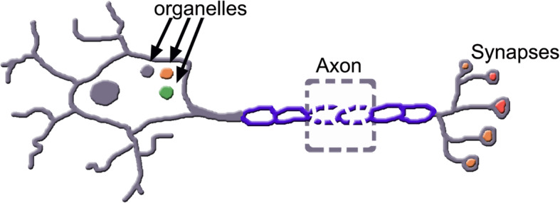
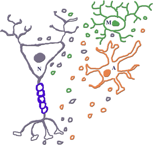
Last century, neurons and glial cells were mostly believed to play distinct functions, relevant for the brain. Progressively, however, it became clear that neurons, astrocytes and microglia co-operate intensely with each other by release/binding of signaling factors, direct surface binding and generation/release of extracellular vesicles, the exosomes and ectosomes, called together vesicles in this abstract. The present review is focused on these vesicles, fundamental in various brain diseases. Their properties are extraordinary. The specificity of their membrane governs their fusion with distinct target cells, variable depending on the state and specificity of their cells of origin and target. Result of vesicle fusion is the discharge of their cargos into the cytoplasm of target cells. Cargos are composed of critical molecules, from proteins (various nature and function) to nucleotides (especially miRNAs), playing critical roles in immune and neurodegenerative diseases. Among immune diseases is multiple sclerosis, affected by extensive dysregulation of co-trafficking neural and glial vesicles, with distinct miRNAs inducing severe or reducing effects. The vesicle-dependent differences between progressive and relapsing-remitting forms of the disease are relevant for clinical developments. In Alzheimer's disease the vesicles can affect the brain by changing their generation and inducing co-release of effective proteins, such Aβ and tau, from neurons and astrocytes. Specific miRNAs can delay the long-term development of the disease. Upon their traffic through the blood-brainbarrier, vesicles of various origin reach fluids where they are essential for the identification of biomarkers, important for diagnostic and therapeutic innovations, critical for the future of many brain patients.
Keywords: Alzheimer’s disease; Astrocytes; Immunological and neurodegenerative diseases; Microglia; Multiple sclerosis; Neurons.
© 2021. The Author(s).
Conflict of interest statement
Not present.
Figures
Similar articles
-
Exosomes and Ectosomes in Intercellular Communication.Curr Biol. 2018 Apr 23;28(8):R435-R444. doi: 10.1016/j.cub.2018.01.059. Curr Biol. 2018. PMID: 29689228 Review.
-
Plasma neuronal exosomes serve as biomarkers of cognitive impairment in HIV infection and Alzheimer's disease.J Neurovirol. 2019 Oct;25(5):702-709. doi: 10.1007/s13365-018-0695-4. Epub 2019 Jan 4. J Neurovirol. 2019. PMID: 30610738 Free PMC article. Review.
-
Neuroinflammation and Depression: Microglia Activation, Extracellular Microvesicles and microRNA Dysregulation.Front Cell Neurosci. 2015 Dec 17;9:476. doi: 10.3389/fncel.2015.00476. eCollection 2015. Front Cell Neurosci. 2015. PMID: 26733805 Free PMC article. Review.
-
Gliocrine System: Astroglia as Secretory Cells of the CNS.Adv Exp Med Biol. 2019;1175:93-115. doi: 10.1007/978-981-13-9913-8_4. Adv Exp Med Biol. 2019. PMID: 31583585 Free PMC article. Review.
-
Extracellular vesicles, news about their role in immune cells: physiology, pathology and diseases.Clin Exp Immunol. 2019 Jun;196(3):318-327. doi: 10.1111/cei.13274. Epub 2019 Mar 11. Clin Exp Immunol. 2019. PMID: 30756386 Free PMC article. Review.
Cited by
-
Modulation of Small RNA Signatures by Astrocytes on Early Neurodegeneration Stages; Implications for Biomarker Discovery.Life (Basel). 2022 Oct 27;12(11):1720. doi: 10.3390/life12111720. Life (Basel). 2022. PMID: 36362875 Free PMC article. Review.
-
Tau and neuroinflammation in Alzheimer's disease: interplay mechanisms and clinical translation.J Neuroinflammation. 2023 Jul 14;20(1):165. doi: 10.1186/s12974-023-02853-3. J Neuroinflammation. 2023. PMID: 37452321 Free PMC article. Review.
-
Four distinct cytoplasmic structures generate and release specific vesicles, thus opening the way to intercellular communication.Extracell Vesicles Circ Nucl Acids. 2023 Mar 15;4(1):44-58. doi: 10.20517/evcna.2023.03. eCollection 2023. Extracell Vesicles Circ Nucl Acids. 2023. PMID: 39698300 Free PMC article. Review.
-
Proteostasis unbalance in prion diseases: Mechanisms of neurodegeneration and therapeutic targets.Front Neurosci. 2022 Sep 6;16:966019. doi: 10.3389/fnins.2022.966019. eCollection 2022. Front Neurosci. 2022. PMID: 36148145 Free PMC article. Review.
-
Neural Circuitry-Related Biomarkers for Drug Development in Psychiatry: An Industry Perspective.Adv Neurobiol. 2024;40:45-65. doi: 10.1007/978-3-031-69491-2_2. Adv Neurobiol. 2024. PMID: 39562440 Review.
References
Publication types
LinkOut - more resources
Full Text Sources
Other Literature Sources