. 2021 Nov 15;2(1):35.
doi: 10.1186/s43556-021-00056-x.

Natural variant frequencies across domains from different sarcomere proteins cross-correlate to identify inter-protein contacts associated with cardiac muscle function and disease

Affiliations

Natural variant frequencies across domains from different sarcomere proteins cross-correlate to identify inter-protein contacts associated with cardiac muscle function and disease

Thomas P Burghardt. Mol Biomed. .

Abstract

Coordinated sarcomere proteins produce contraction force for muscle shortening. In human ventriculum they include the cardiac myosin motor (βmys), repetitively converting ATP free energy into work, and myosin binding protein C (MYBPC3) that in complex with βmys is regulatory. Single nucleotide variants (SNVs) causing hereditary heart diseases frequently target this protein pair. The βmys/MYBPC3 complex models a regulated motor and is used here to study how the proteins couple. SNVs in βmys or MYBPC3 survey human populations worldwide. Their protein expression modifies domain structure affecting phenotype and pathogenicity outcomes. When the SNV modified domain locates to inter-protein contacts it could affect complex coordination. Domains involved, one in βmys the other in MYBPC3, form coordinated domains (co-domains). Co-domain bilateral structure implies the possibility for a shared impact from SNV modification in either domain suggesting a correlated response to a common perturbation could identify their location. Genetic divergence over human populations is proposed to perturb SNV probability coupling that is detected by cross-correlation in 2D correlation genetics (2D-CG). SNV probability data and 2D-CG identify three critical sites, two in MYBPC3 with links to several domains across the βmys motor, and, one in βmys with links to the MYBPC3 regulatory domain. MYBPC3 sites are hinges sterically enabling regulatory interactions with βmys. The βmys site is the actin binding C-loop (residues 359-377). The C-loop is a trigger for actin-activated myosin ATPase and a contraction velocity modulator. Co-domain identification implies their spatial proximity suggesting a novel approach for in vivo protein complex structure determination.

Keywords: 2D Correlation genetics; Cardiac muscle inheritable disease; Interprotein coordinated domains; Population genetic divergence proxy; Serial founder effect; Single nucleotide variants.


Conflict of interest statement

The author has no relevant financial or non-financial interest to disclose.

Figures

Fig. 1.
Fig. 1.
Myosin and MYBPC3 structures in the sarcomere. Myosin dimer model (bottom right) has two subfragment 1 (SF1), subfragment 2 (S2), and light meromyosin (LM) polypeptides. SF1 crystal structure has a motor domain and a lever arm (blue) with bound light chains ELC (black) and RLC (red). The motor domain contains several binding sites for actin, the active site binding ATP (green), the switch 2 helix (blue), SH1/SH2 hinge (silver), and converter domain (red). MYBPC3 model (upper right) has multiple domains shown schematically in black circles or red squares indicating immunoglobulin-like or fibronectin-like domains with CX binding myosin LM, N-terminal C0-C2 maintaining transient interactions with F-actin (green) and myosin, and the linker containing regulatory phosphorylation sites. Tension and load vectors indicate direction of force generated by myosin and opposed by F-actin, respectively. F-actin and SF1 homology models were obtained as described [1]
Fig. 2.
Fig. 2.
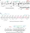
Linearized diagrams for βmys, MYBPC3, and microtubule-associated protein tau (MAPT). Diagrams identify most domains defined in Supplementary Tables S1 and S4 and approximate residue numbering. a The βmys diagram has a heavy chain (H3 through LM), ELC (EN through E3), and RLC (RN through R3). Heavy chain diagram does not indicate the active site (AC), Omecamtiv Mecarbil binding site (OM), and mesa (ME) because they occupy multiple regions in the linear representation. MYBPC3 and actin binding sites are indicated in red and green above the heavy chain and below the light chains. b MYBPC3 has Ig-like (black circles) and fibronectin-like domains (red squares). Serine phosphorylation sites, S1 (Ser47), ST (Ser273), S3 (Ser282), S4 (Ser302), and S5-S7 and a threonine phosphorylation site (S8) are indicated below the chain. Domain linkers of interest include the proline rich linker (PR) and LT containing a regulatory site. Z1 is a zinc binding site. Myosin RLC, S2, LM, and actin binding sites on MYBPC3 are indicated above the linearized model. c The MAPT diagram shows the largest isoform (isoform 1). Sites or domains identified are tyrosine phosphorylation sites (y1 and y2), N-terminal inserts (n1 and n2), proline rich regions (p1-p3), microtubule binding repeats (t1-t4), cysteines C608 and C639 (s9 and s2), and hexapeptide motifs PHF6* (VQIINK at x1) and PHF6 (VQIVYK at x2)
Fig. 3
Fig. 3
Hypothetical βmys/MYBPC3 complexes in human populations for SNVs detecting real and virtual co-domains. Functional domains CL and ML in βmys, and, functional domains S3 and S7 in MYBPC3 are SNV modified individually as shown in a, c, b, and d. Complexes SNV (red star) modified at C-loop (a), phosphorylatable serine S3 (b), myopathy loop (c), or phosphorylatable serine S7 (d) are species needed in the database to surmise the co-domain interactions in the native species shown in f. The native complex involves real co-domains indicated by the solid green lines between S3 and CL and between S7 and ML. Co-domain interruption by SNVs is indicated by the solid red lines connecting S3-CL or S7-ML in a-d. SNV modification in e locates to L8 (red triangle) where it alters MYBPC3 conformation to interrupt real co-domains S3-CL and S7-ML. The virtual co-domains L8-CL and L8-ML are indicated by the broken green lines in f
Fig. 4
Fig. 4
βmys/MYBPC3 complex correlation maps for pathogenic outcomes implied by 2D-CG. Synchronous (left) or asynchronous (right) maps have axes identical for both dimensions representing βmys (M7 1-39) followed by MYBPC3 (C3 40-65) domains. Domain index is linked to its two-letter code and protein sequence in Supplementary Table S1. Two-letter codes for domains label some indices on the leftmost axis in the figure. Intensities (z-values) are indicated numerically by the grayscale. Regions defined by vertical and horizontal orange lines at the interface of pixels 39-40 separate intra-protein cross-peaks (within regions M7-M7 and C3-C3) from inter-protein cross-peaks (within regions M7-C3 and C3-M7). Intensity peaks along the diagonal in the synchronous map are autocorrelated probabilities for each domain. Correlation squares link the 6 most significant off-diagonal co-domain coordinates falling within the M7-C3 and C3-M7 regions. They are white, green, red, blue, for different βmys domains then repeating color sequence as needed
Fig. 5
Fig. 5
βmys/MYBPC3 complex cross-correlation maps and their implied inter-protein domain linkages for pathogenic outcomes. a M7-C3 region for synchronous and asynchronous maps and the portion of the correlation squares falling within them for pathogenic outcomes taken from Fig. 4. Co-domain ordered pairs corresponding to each correlation square are indicated in parenthesis for M7 domain and C3 domain in first and second positions, respectively. Ordered pairs are color coded to match the corresponding correlation square. b Red lines show most significant directed co-domain interactions between βmys and MYBPC3 for pathogenic outcomes. Nomenclature otherwise identical to Fig. 4
Fig. 6.
Fig. 6.
βmys/MYBPC3 complex cross-correlation maps and their implied inter-protein domain linkages for benign outcomes. a M7-C3 region for synchronous and asynchronous maps and the portion of the correlation squares falling within them for benign outcomes taken from Supplementary Figure S4. b Red lines show most significant directed co-domain interactions between βmys and MYBPC3 for benign outcomes. Nomenclature otherwise identical to Figs. 4 and 5
Fig. 7.
Fig. 7.
MAPT/MYBPC2 complex cross-correlation maps and their implied inter-protein domain linkages for pathogenic outcomes. a C3-MT region for synchronous and asynchronous maps and the portion of the correlation squares falling within them for pathogenic outcomes taken from Supplementary Figure S5. b Red lines show most significant directed co-domain interactions between MAPT and MYBPC3 for pathogenic outcomes. Nomenclature otherwise identical to Figs. 4 and 5
Fig. 8.
Fig. 8.
MAPT/MYBPC2 complex cross-correlation maps and their implied inter-protein domain linkages for benign outcomes. a C3-MT region for synchronous and asynchronous maps and the portion of the correlation squares falling within them for benign outcomes taken from Supplementary Figure S6. b Red lines show most significant directed co-domain interactions between MAPT and MYBPC3 for benign outcomes. Nomenclature is otherwise identical to Figs. 4 and 5

Similar articles

Cited by

References

    1. Wang Y, Ajtai K, Burghardt Thomas P. Cardiac and skeletal actin substrates uniquely tune cardiac myosin strain-dependent mechanics. Open Biol. 2018;8(11):180143. doi: 10.1098/rsob.180143. - DOI - PMC - PubMed
    1. Al-Khayat HA, Kensler RW, Squire JM, Marston SB, Morris EP. Atomic model of the human cardiac muscle myosin filament. Proc Natl Acad Sci USA. 2013;110(1):318–323. doi: 10.1073/pnas.1212708110. - DOI - PMC - PubMed
    1. Lorenz M, Holmes KC. The actin-myosin interface. Proc Natl Acad Sci U S A. 2010;107:12529–12534. doi: 10.1073/pnas.1003604107. - DOI - PMC - PubMed
    1. Highsmith S, Eden D. Myosin-ATP chemomechanics. Biochemistry. 1993;32:2455–2458. doi: 10.1021/bi00061a001. - DOI - PubMed
    1. Rayment I, Holden HM. Myosin subfragment-1: structure and function of a molecular motor. Curr Opin Struct Biol. 1993;1993:944–952. doi: 10.1016/0959-440X(93)90160-M. - DOI

LinkOut - more resources