Review
. 2021 Jul 30;2(1):23.
doi: 10.1186/s43556-021-00043-2.

E3 ubiquitin ligases: styles, structures and functions

Affiliations
Review

E3 ubiquitin ligases: styles, structures and functions

Quan Yang et al. Mol Biomed. .

Abstract

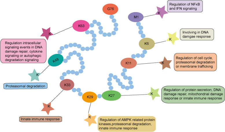
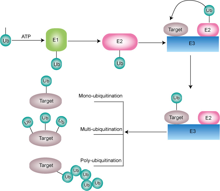
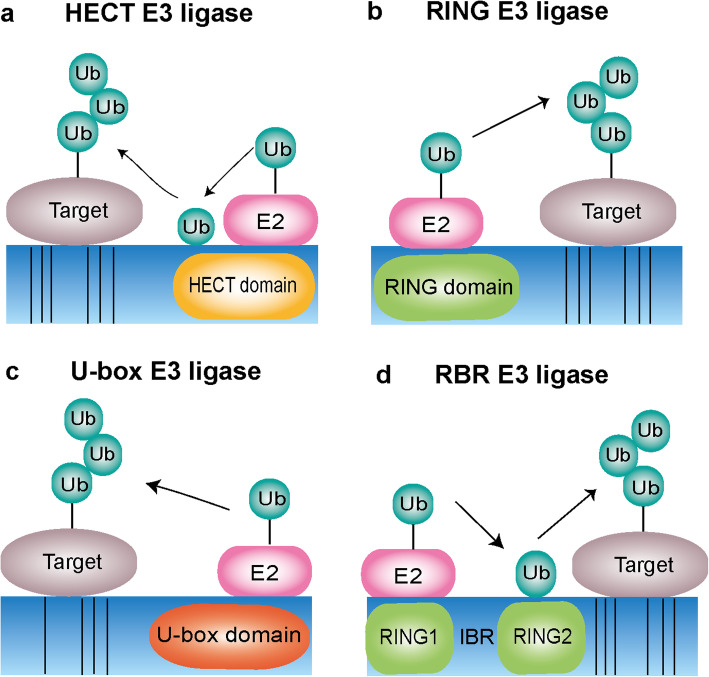
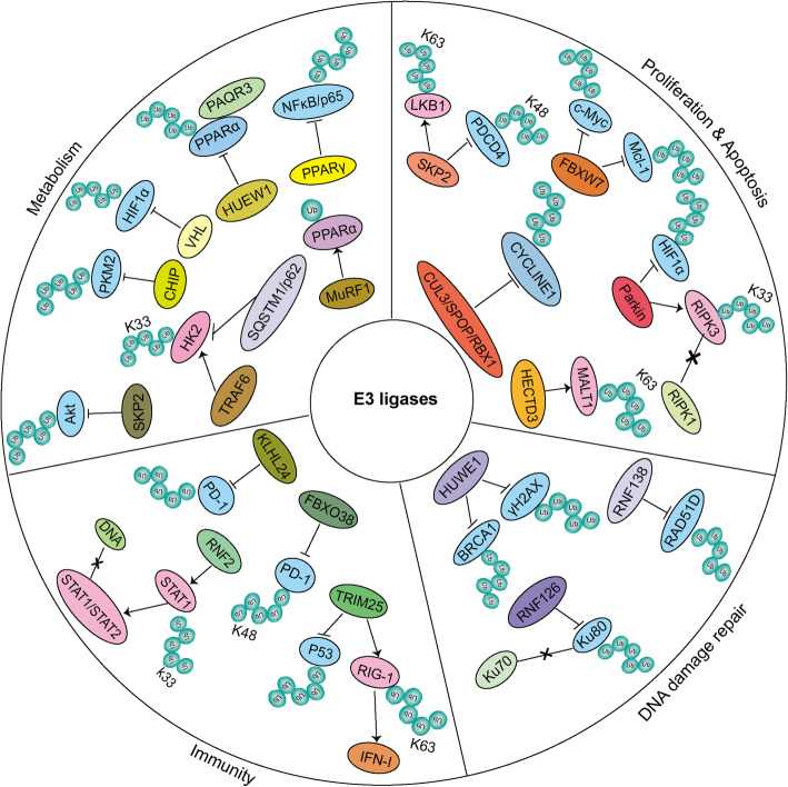
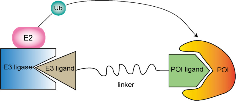
E3 ubiquitin ligases are a large family of enzymes that join in a three-enzyme ubiquitination cascade together with ubiquitin activating enzyme E1 and ubiquitin conjugating enzyme E2. E3 ubiquitin ligases play an essential role in catalyzing the ubiquitination process and transferring ubiquitin protein to attach the lysine site of targeted substrates. Importantly, ubiquitination modification is involved in almost all life activities of eukaryotes. Thus, E3 ligases might be involved in regulating various biological processes and cellular responses to stress signal associated with cancer development. Thanks to their multi-functions, E3 ligases can be a promising target of cancer therapy. A deeper understanding of the regulatory mechanisms of E3 ligases in tumorigenesis will help to find new prognostic markers and accelerate the growth of anticancer therapeutic approaches. In general, we mainly introduce the classifications of E3 ligases and their important roles in cancer progression and therapeutic functions.

Keywords: 26S proteasome degradation; Cancer progression; E3 ligases; PROTACs; Therapeutics; Ubiquitination.


Conflict of interest statement

The authors have declared that no competing interest exists.

Figures

Fig. 1
Fig. 1
Different ubiquitin linkage types and their functions. Ubiquitin (Ub) a small protein consists of 76 amino acids highly conserved among all eukaryotes. Ubiquitin is characterized by its 7 lysine sites (K6, K11, K27, K29, K33, K48, K63) and N-terminal methionine1 (Met1) site, which are also functional sites. Due to these specific linkage sites, ubiquitination can be classified into different styles and then perform distinct biological functions. And ubiquitin attaches to target substrate through identifying its lysine (K) site
Fig. 2
Fig. 2
Overview of the cascade process of ubiquitination. Ubiquitination is an important cascade process of posttranslational modification catalyzed by three key enzymes. Firstly, E1 catalyzes the activation of Ub through a thioester bond in an ATP dependent mechanism. Then, activated Ub is transferred to the active-site cysteine residue of an E2. The last step is mediated by an E3 ligase that recognizes the E2 complex and facilitates the transfer of Ub from E2 to the target substrate. Due to the different binding style of Ub on the target substrate, types of ubiquitination modification are divided into three: mono-ubiquitination, multi-ubiquitination and poly-ubiquitination
Fig. 3
Fig. 3
Types of ubiquitination ligases. a The HECT type E3 ligases contain the conserved C-terminal HECT domain and the N-terminal consists with different domains depending the specific subtype. HECT type E3 ligases involved ubiquitination process including a two-step reaction: ubiquitin is first carried by E2 ligase binding to the HECT domain and then transferred to a catalytic cysteine on the E3 ligase, the second step is the transfer of ubiquitin from the E3 ligase to the substrate. b The RING type E3 ligases are characterized by the presence of a zinc-binding domain called RING at the N-terminal. RING E3s mediate a direct transfer of ubiquitin from E2 ligase to the substrate. c The U-box type E3 ligases contain U-box domain at the C-terminal which is responsible for binding the ubiquitin-charged E2 ligase and stimulating ubiquitin transfer. d The RBR type E3 ligases consist of two predicted RING domains (RING1 and RING2) separated by IBR domain . RBR E3 ligases catalyzed ubiquitination process involves a two-step reaction where ubiquitin is first transferred to a catalytic RING2 domain on the E3 and then to the substrate
Fig. 4
Fig. 4
Multi-functions of E3 ligases. Based on the different E3 ligases and their specific substrates, E3 ligases can involve in many different cellular progression such as proliferation, apoptosis, DNA damage repair, immunity and metabolism. We have shown some E3 ligases mentioned in this review and how they function in regulating cancer cell progression. For example, the E3 ligases FBXW7, HECTD3, CUL3/SPOP/RBX1, Parkin, SKP2 are involved in regulating proliferation or apoptosis through targeting proliferation-associated proteins for ubiquitination. E3 ligases HUWE1, RNF126, RNF138 are shown to regulate cancer cell DNA damage repair by targeting specific substrates for ubiquitination. E3 ligases FBXO38, KLHL22, TRIM25, RNF2 regulate cancer cell immune response through promoting ubiquitination of specific substrates. And E3 ligases PPARγ, SKP2, CHIP, VHL, TRAF6, HUEW1,MuRF1 are capable of mediating cancer cell metabolism by targeting related substrates for ubiquitination. Note:we use different background colors to distinguish each E3 ligases. Blue background represents the genes they target for degradation and pink background represnets genes they target for stabilization or others but not degradation. We use × to show the disruption of protein-protein interaction
Fig. 5
Fig. 5
Schematic diagram of PROTACs. A Graphical representation of the components and process of PROTACs. The PROTACs consist of three important parts, including a ligand binding to the POI, a covalently linked ligand of an E3 ubiquitin ligase and a linker to link these two ligands

References

    1. Dikic I. Proteasomal and autophagic degradation systems. Annu Rev Biochem. 2017;86:193–224. doi: 10.1146/annurev-biochem-061516-044908. - DOI - PubMed
    1. Lamb CA, Yoshimori T, Tooze SA. The autophagosome: origins unknown, biogenesis complex. Nat Rev Mol Cell Biol. 2013;14(12):759–774. doi: 10.1038/nrm3696. - DOI - PubMed
    1. Finley D. Recognition and processing of ubiquitin-protein conjugates by the proteasome. Annu RevBiochem. 2009;78:477–513. doi: 10.1146/annurev.biochem.78.081507.101607. - DOI - PMC - PubMed
    1. Tokheim C, Wang X, Timms RT, Zhang B, Mena EL, Wang B, et al. Systematic characterization of mutations altering protein degradation in human cancers. Mol Cell. 2021. 10.1016/j.molcel.2021.01.020. - PMC - PubMed
    1. Goldberg AL, Stein R, Adams J. New insights into proteasome function: from archaebacteria to drug development. Chem Biol. 1995;2(8):503–508. doi: 10.1016/1074-5521(95)90182-5. - DOI - PubMed

LinkOut - more resources