. 2022 Aug;76(2):372-386.
doi: 10.1002/hep.32326. Epub 2022 Mar 4.

Progressive loss of hepatocyte nuclear factor 4 alpha activity in chronic liver diseases in humans

Affiliations

Progressive loss of hepatocyte nuclear factor 4 alpha activity in chronic liver diseases in humans

Sumedha Gunewardena et al. Hepatology. 2022 Aug.

Abstract

Background and aims: Hepatocyte nuclear factor 4 alpha (HNF4α) is indispensable for hepatocyte differentiation and critical for maintaining liver health. Here, we demonstrate that loss of HNF4α activity is a crucial step in the pathogenesis of chronic liver diseases (CLDs) that lead to development of HCC.

Approach and results: We developed an HNF4α target gene signature, which can accurately determine HNF4α activity, and performed an exhaustive in silico analysis using hierarchical and K-means clustering, survival, and rank-order analysis of 30 independent data sets containing over 3500 individual samples. The association of changes in HNF4α activity to CLD progression of various etiologies, including HCV- and HBV-induced liver cirrhosis (LC), NAFLD/NASH, and HCC, was determined. Results revealed a step-wise reduction in HNF4α activity with each progressive stage of pathogenesis. Cluster analysis of LC gene expression data sets using the HNF4α signature showed that loss of HNF4α activity was associated with progression of Child-Pugh class, faster decompensation, incidence of HCC, and lower survival with and without HCC. A moderate decrease in HNF4α activity was observed in NAFLD from normal liver, but a further significant decline was observed in patients from NAFLD to NASH. In HCC, loss of HNF4α activity was associated with advanced disease, increased inflammatory changes, portal vein thrombosis, and substantially lower survival.

Conclusions: In conclusion, these data indicate that loss of HNF4α function is a common event in the pathogenesis of CLDs leading to HCC and is important from both diagnostic and therapeutic standpoints.


Conflict of interest statement

Conflict of interest statement:

None of the authors have any conflict to declare

Figures

Figure 1.
Figure 1.
Development and validation of HNF4α target gene signature to determine HNF4α function. Scheme shows the experimental design of the bioinformatics analysis to identify the HNF4α target gene signature.
Figure 2.
Figure 2.
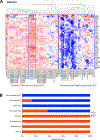
Progressive loss of HNF4α function during various stages of HCC pathogenesis. (A) HNF4α target gene signature was used to perform cluster analysis on global gene expression data from GSE6764, a dataset containing 76 individual samples of normal livers and various pathological stages including low- and high-grade dysplasia, cirrhosis, early stage and advanced HCC. Cluster analysis yielded two main clusters. C1, outlined by solid red line, mainly included normal and cirrhotic samples, and C2, outlined by solid blue line, contained mainly advanced and highly advanced HCC. Further, two subclusters (SC3 and SC4) were observed showing normal and very advanced HCC, respectively. White color in the heatmaps represents no change (no difference), blue color represents decreased (lower) expression and red represents increased (higher) expression. (B) Bar graph showing percent of each type of cases with either C1 or C2.
Figure 3.
Figure 3.
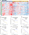
Loss of HNF4α activity is a critical determinant of cirrhosis outcome. Cluster analysis was performed on dataset GSE15654 containing gene expression data from 216 early stage (Child-Pugh class A) liver cirrhosis samples. Further, Kaplan-Meier (K-M) survival estimation analysis was conducted to determine how each of the clusters performed for various endpoint. (A) Heatmap showing cluster analysis. White color in the heatmaps represents no change (no difference), blue color represents decreased (lower) expression and red represents increased (higher) expression. K-M plots were generated to determine probability of the two groups (group 1 with high HNF4α activity and group 2 with low HNF4α activity) for reaching various clinical endpoints including (B) change (increase) in cirrhosis stage, (C) decompensation, (D) development of HCC, (E) overall survival, (F) survival in cases which did not develop HCC, and (G) survival in cases that developed HCC.
Figure 4.
Figure 4.
Significant loss of HNF4α activity in nonalcoholic steatohepatitis (NASH). (A) Cluster analysis using the HNF4α target gene signature performed on dataset GSE 33814 containing 44 samples of normal, steatosis and steatohepatitis (NASH) patients revealed three clusters. Cluster 1 contained 80% normal and 20% steatosis samples, cluster 2 contained 20% normal, 60% steatosis and 20% NASH, and cluster 3 contained 1% normal, 35% steatosis and 57% NASH samples. Rank order analysis of samples from dataset GSE135251 containing 20 NAFLD/NASH and 10 control samples according to various clinical parameters including (B) NASH stage, (C) NAS score, (D) fibrosis score, and (E) early vs advanced stage of NASH.
Figure 5.
Figure 5.
HCC shows significant loss of HNF4α activity as compared to surrounding normal tissue. Cluster analysis using HNF4α target gene signature was performed on datasets containing gene expression data from paired tumor and surrounding non-tumor normal (surrounding NT) tissues. (A) dataset GSE60502 consisting of 36 samples (18 pairs), (B) dataset GSE14520-GLP571 containing 38 samples (19 pairs), (C) dataset GSE14520-GLP3921 containing 426 samples (213 pairs), (D) dataset GSE64041 containing 125 samples (60 pairs plus 5 normal tissues) of HCC and surrounding normal tissues. White color in the heatmaps represents no change (no difference), blue color represents decreased (lower) expression and red represents increased (higher) expression.
Figure 6.
Figure 6.
HCC patients with higher HNF4α activity have higher survival. Cluster analysis using HNF4α target gene signature was performed on two datasets and accompanied survival data was used for Kaplan-Meier survival estimation analysis (K-M) (A), and (C) show heatmaps obtained from cluster analysis and (B), (D) show K-M plot of survival data from TCGA dataset and dataset GSE10143, respectively. (E) shows K-M analysis of HCC recurrence for dataset GSE10143. For (A) black color represents no change, red color represents increased (higher) gene expression and green color shows decreased (lower) gene expression. For (C) white color represents no change (no difference), blue color represents decreased (lower) expression and red represents increased (higher) expression.
Figure 7.
Figure 7.
Rank order analysis using HNF4α activity as measured by HNF4α target gene signature expression shows stepwise decline in HNF4α during chronic liver diseases. Rank order graphs of (A) dataset GSE62232 dataset containing 81 pairs of tumors and surrounding normal tissues separated by Edmondson Grades I to IV, (B) dataset GSE84044 dataset consisting of 124 HBV-associated liver cirrhosis separated by Scheurer score for inflammation, and (C) of the same data set separated by Scheurer score for fibrosis

Similar articles

Cited by

References

    1. Gonzalez FJ. Regulation of hepatocyte nuclear factor 4 alpha-mediated transcription. Drug Metab Pharmacokinet 2008;23:2–7. - PubMed
    1. Sladek FM. Orphan receptor HNF-4 and liver-specific gene expression. Receptor 1994;4:64. - PubMed
    1. Watt AJ, Garrison WD, Duncan SA. HNF4: a central regulator of hepatocyte differentiation and function. Hepatology 2003;37:1249–1253. - PubMed
    1. Walesky C, Apte U. Role of hepatocyte nuclear factor 4alpha (HNF4alpha) in cell proliferation and cancer. Gene Expr 2015;16:101–108. - PMC - PubMed
    1. Li J, Ning G, Duncan SA. Mammalian hepatocyte differentiation requires the transcription factor HNF-4alpha. Genes Dev 2000;14:464–474. - PMC - PubMed

Publication types

MeSH terms

Substances