

Cannabinoids Block Cellular Entry of SARS-CoV-2 and the Emerging Variants
- PMID: 35007072
- PMCID: PMC8768006
- DOI: 10.1021/acs.jnatprod.1c00946
Cannabinoids Block Cellular Entry of SARS-CoV-2 and the Emerging Variants
Abstract
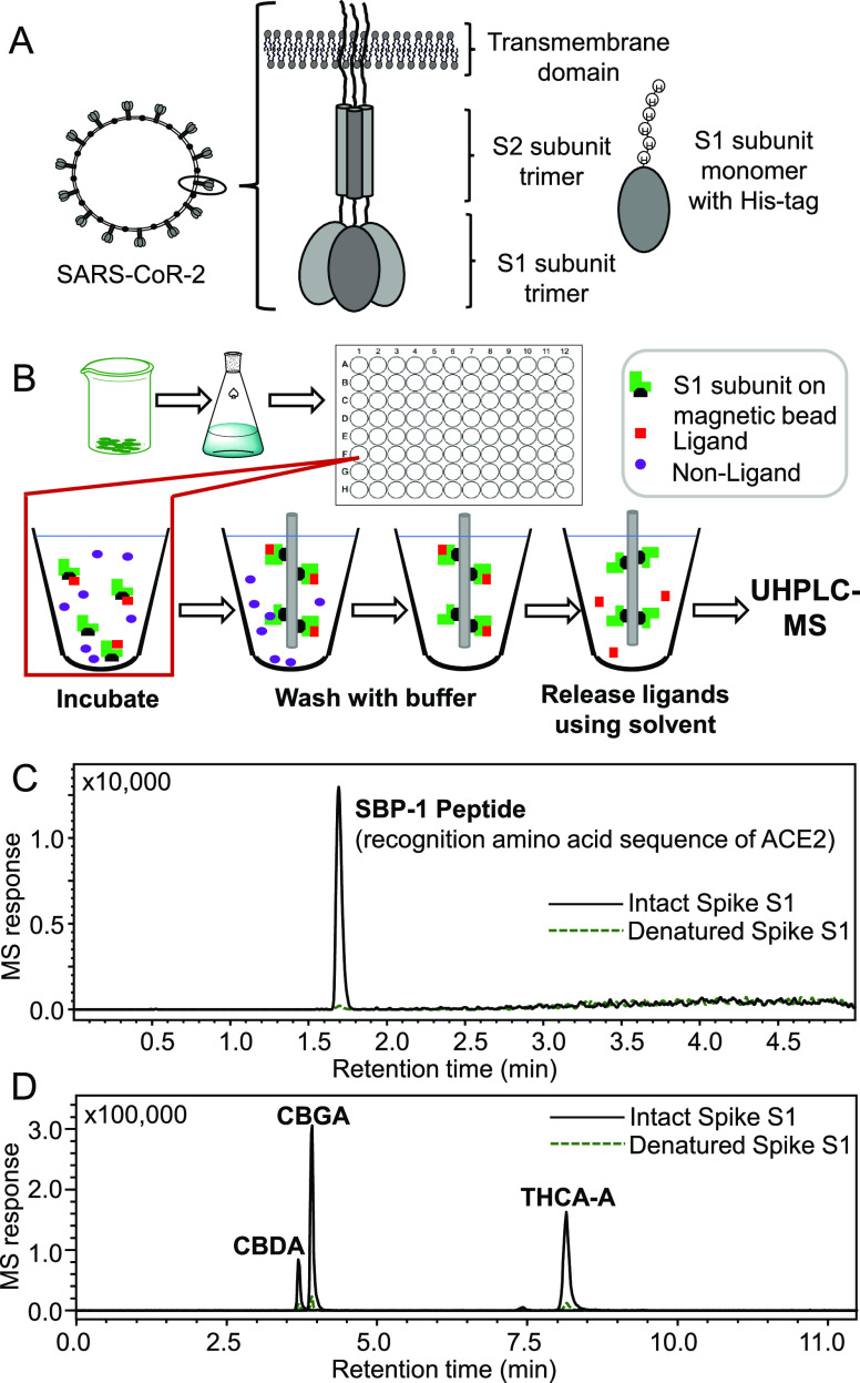
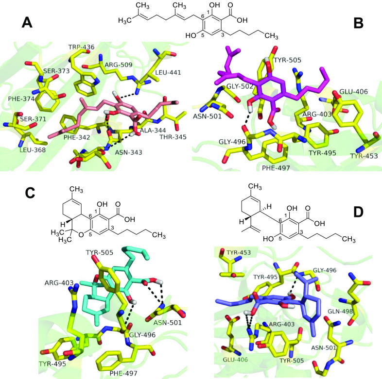
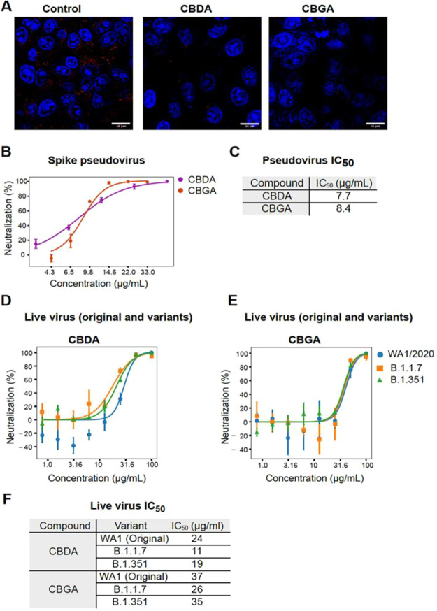
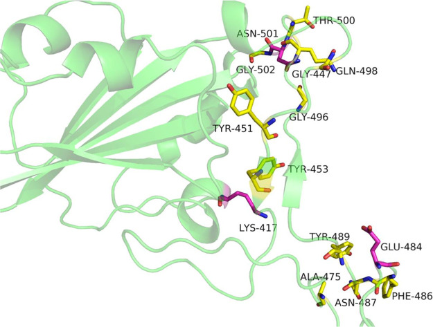
As a complement to vaccines, small-molecule therapeutic agents are needed to treat or prevent infections by severe acute respiratory syndrome coronavirus-2 (SARS-CoV-2) and its variants, which cause COVID-19. Affinity selection-mass spectrometry was used for the discovery of botanical ligands to the SARS-CoV-2 spike protein. Cannabinoid acids from hemp (Cannabis sativa) were found to be allosteric as well as orthosteric ligands with micromolar affinity for the spike protein. In follow-up virus neutralization assays, cannabigerolic acid and cannabidiolic acid prevented infection of human epithelial cells by a pseudovirus expressing the SARS-CoV-2 spike protein and prevented entry of live SARS-CoV-2 into cells. Importantly, cannabigerolic acid and cannabidiolic acid were equally effective against the SARS-CoV-2 alpha variant B.1.1.7 and the beta variant B.1.351. Orally bioavailable and with a long history of safe human use, these cannabinoids, isolated or in hemp extracts, have the potential to prevent as well as treat infection by SARS-CoV-2.
Conflict of interest statement
The authors declare no competing financial interest.
Figures
Similar articles
-
Discovery and Evaluation of Entry Inhibitors for SARS-CoV-2 and Its Emerging Variants.J Virol. 2021 Nov 23;95(24):e0143721. doi: 10.1128/JVI.01437-21. Epub 2021 Sep 22. J Virol. 2021. PMID: 34550770 Free PMC article.
-
Inhibition of ACE2-Spike Interaction by an ACE2 Binder Suppresses SARS-CoV-2 Entry.Angew Chem Int Ed Engl. 2022 Mar 7;61(11):e202115695. doi: 10.1002/anie.202115695. Epub 2022 Feb 1. Angew Chem Int Ed Engl. 2022. PMID: 35043545 Free PMC article.
-
Identification of nitrile-containing isoquinoline-related natural product derivatives as coronavirus entry inhibitors in silico and in vitro.Biomed Pharmacother. 2024 Nov;180:117517. doi: 10.1016/j.biopha.2024.117517. Epub 2024 Oct 1. Biomed Pharmacother. 2024. PMID: 39357326
-
Inhibition of S-protein RBD and hACE2 Interaction for Control of SARSCoV- 2 Infection (COVID-19).Mini Rev Med Chem. 2021;21(6):689-703. doi: 10.2174/1389557520666201117111259. Mini Rev Med Chem. 2021. PMID: 33208074 Review.
-
Potential therapeutic approaches for the early entry of SARS-CoV-2 by interrupting the interaction between the spike protein on SARS-CoV-2 and angiotensin-converting enzyme 2 (ACE2).Biochem Pharmacol. 2021 Oct;192:114724. doi: 10.1016/j.bcp.2021.114724. Epub 2021 Aug 8. Biochem Pharmacol. 2021. PMID: 34371003 Free PMC article. Review.
Cited by
-
Identification of SARS-CoV-2 Main Protease Inhibitors from a Library of Minor Cannabinoids by Biochemical Inhibition Assay and Surface Plasmon Resonance Characterized Binding Affinity.Molecules. 2022 Sep 19;27(18):6127. doi: 10.3390/molecules27186127. Molecules. 2022. PMID: 36144858 Free PMC article.
-
Anti-Inflammatory and Antiviral Effects of Cannabinoids in Inhibiting and Preventing SARS-CoV-2 Infection.Int J Mol Sci. 2022 Apr 10;23(8):4170. doi: 10.3390/ijms23084170. Int J Mol Sci. 2022. PMID: 35456990 Free PMC article. Review.
-
Evaluating the Metabolomic Profile and Anti-Pathogenic Properties of Cannabis Species.Metabolites. 2024 Apr 26;14(5):253. doi: 10.3390/metabo14050253. Metabolites. 2024. PMID: 38786730 Free PMC article. Review.
-
Molecular Docking and Dynamics Simulation of Several Flavonoids Predict Cyanidin as an Effective Drug Candidate against SARS-CoV-2 Spike Protein.Adv Pharmacol Pharm Sci. 2022 Nov 9;2022:3742318. doi: 10.1155/2022/3742318. eCollection 2022. Adv Pharmacol Pharm Sci. 2022. PMID: 36407836 Free PMC article.
-
Therapeutic Potential of Cannabis: A Comprehensive Review of Current and Future Applications.Biomedicines. 2023 Sep 25;11(10):2630. doi: 10.3390/biomedicines11102630. Biomedicines. 2023. PMID: 37893004 Free PMC article. Review.
References
-
- https://www.worldometers.info/coronavirus/, accessed 15 Dec 2021.
-
- Centers for Disease Control and Prevention. https://www.cdc.gov/coronavirus/2019-ncov/science/science-briefs/scienti..., accessed 15 Dec 2021.
-
- Huang C.; Wang Y.; Li X.; Ren L.; Zhao J.; Hu Y.; Zhang L.; Fan G.; Xu J.; Gu X.; Cheng Z.; Yu T.; Xia J.; Wei Y.; Wu W.; Xie X.; Yin W.; Li H.; Liu M.; Xiao Y.; Gao H.; Guo L.; Xie J.; Wang G.; Jiang R.; Gao Z.; Jin Q.; Wang J.; Cao B. Lancet 2020, 395, 497–506. 10.1016/S0140-6736(20)30183-5. - DOI - PMC - PubMed
MeSH terms
Substances
Supplementary concepts
Grants and funding
LinkOut - more resources
Full Text Sources
Other Literature Sources
Research Materials
Miscellaneous
