Review
. 2021 Dec 24;14(1):74.
doi: 10.3390/cancers14010074.

Diagnosis of Glioblastoma by Immuno-Positron Emission Tomography

Affiliations
Review

Diagnosis of Glioblastoma by Immuno-Positron Emission Tomography

Eduardo Ruiz-López et al. Cancers (Basel). .

Abstract

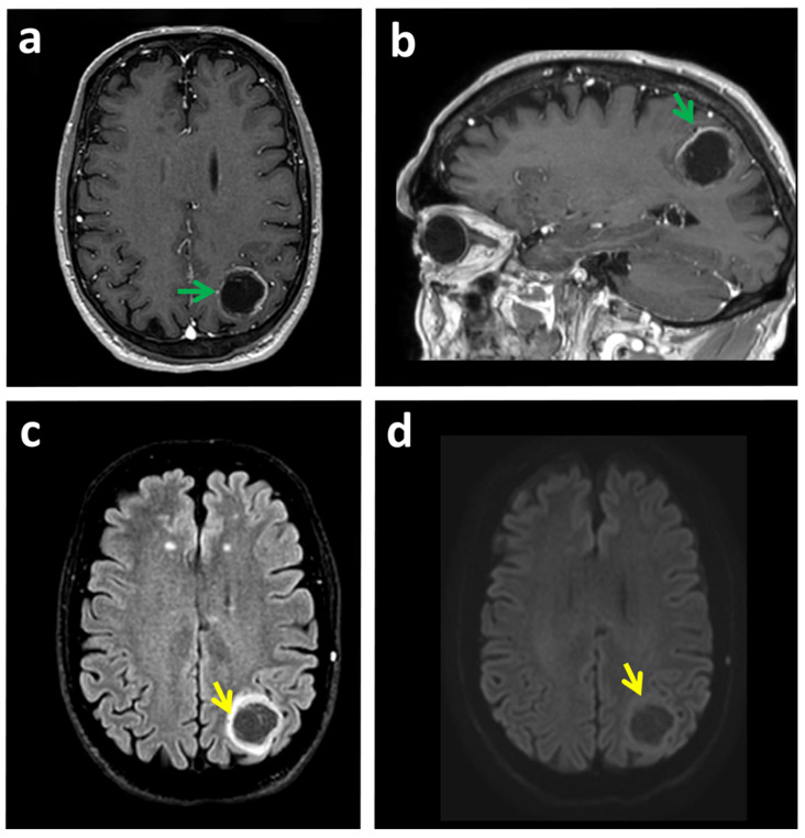
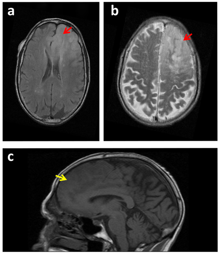
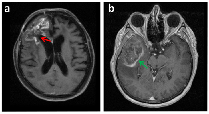
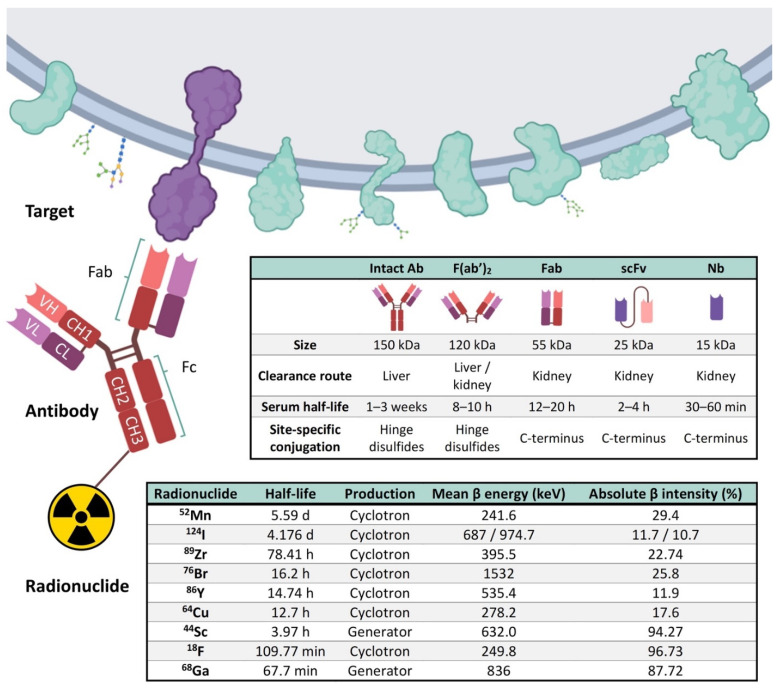
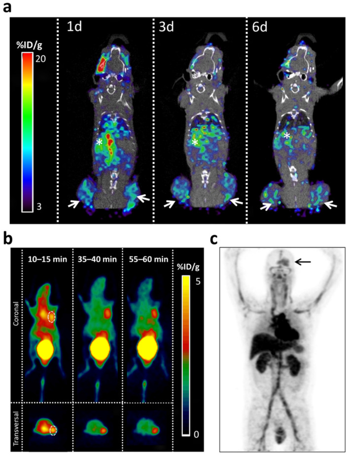
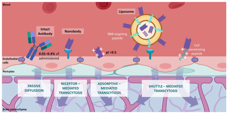
Neuroimaging has transformed neuro-oncology and the way that glioblastoma is diagnosed and treated. Magnetic Resonance Imaging (MRI) is the most widely used non-invasive technique in the primary diagnosis of glioblastoma. Although MRI provides very powerful anatomical information, it has proven to be of limited value for diagnosing glioblastomas in some situations. The final diagnosis requires a brain biopsy that may not depict the high intratumoral heterogeneity present in this tumor type. The revolution in "cancer-omics" is transforming the molecular classification of gliomas. However, many of the clinically relevant alterations revealed by these studies have not yet been integrated into the clinical management of patients, in part due to the lack of non-invasive biomarker-based imaging tools. An innovative option for biomarker identification in vivo is termed "immunotargeted imaging". By merging the high target specificity of antibodies with the high spatial resolution, sensitivity, and quantitative capabilities of positron emission tomography (PET), "Immuno-PET" allows us to conduct the non-invasive diagnosis and monitoring of patients over time using antibody-based probes as an in vivo, integrated, quantifiable, 3D, full-body "immunohistochemistry" in patients. This review provides the state of the art of immuno-PET applications and future perspectives on this imaging approach for glioblastoma.

Keywords: antibody; diagnostic imaging; glioblastoma; immuno-PET; molecular imaging; nanobody; neuroimaging; theragnostic probes.


Conflict of interest statement

The authors declare no conflict of interest.

Figures

Figure 1
Figure 1
A case of glioma that could be confounded with brain access by MRI. (a,b) MRI images of a patient with glioblastoma in the left parieto-occipital lobe. T1W_3D-FFE MRI with gadolinium paramagnetic contrast. (a) Axial and (b) Sagittal reconstruction. The tumor shows contrast rim-enhancement (green arrow). This lesion was confounded with a brain abscess. (c) Fluid-attenuated inversion recovery (FLAIR) shows a parieto-occipital space-occupying lesion with peripheral hyperintensity and central hypointensity (yellow arrow). (d) The diffusion sequence shows minimal restriction of hydric diffusion (yellow arrow), which excludes the possibility that it is an abscess with typical behavior. Biopsy confirmed a diagnosis of glioblastoma.
Figure 2
Figure 2
MRI scans of a case of glioma that could be confounded with an ischemic stroke. (ac) MRI images of a patient with a glioma in the right frontal lobe (red arrows). (a) Inversion recovery fast spin-echo (IRFSE) Fluid-Attenuated Inversion Recovery (FLAIR), axial MRI. (b) Axial FSE T2 MRI image. (c) Spin-echo (SE) T1 sagittal MRI image. The space-occupying lesion could be confounded with an ischemic stroke in evolution (yellow arrows). Loss of gray and white matter differentiation. The lesion was confirmed to be a diffuse tumoral mass compatible with grade II astrocytoma by anatomopathological analysis.
Figure 3
Figure 3
Other lesions can be confounding with glioblastoma. (a) Axial FS T1 MRI image with contrast of a glioblastoma recurrence (red arrow). In some situations, conventional MRI cannot correctly differentiate tumor tissue from post-therapeutic effects following neurosurgical resection and radiation. In this image, tumor recurrence was confounded with treatment necrosis produced by radiation. (b) Axial 3D Fast spoiled gradient echo (FSPGR) with MRI image. A patient suffering from hepatocellular carcinoma (HCC) presented one brain lesion detected by MRI (green arrow). In this situation a glioblastoma could be confounding with a brain metastasis. A biopsy indicated a glioblastoma and was discarded to be a brain metastasis from the HCC.
Figure 4
Figure 4
Representation of the three main components of the immuno-PET. Targets present in the external surface of the plasma membrane, antibody, and its derived immune fragments F(ab′)2, Fab, scFv, and Nb, and the most commonly used radionuclides are represented. A typical antibody (Immunoglobulin G, IgG) is composed of two heavy (H) chains and 2 light (L) chains. Heavy chains contain a series of immunoglobulin domains, usually with one variable domain (VH) that is important for antigen binding, and several constant domains (CH1, CH2, CH3). Light chains are composed of one variable (VL) and one constant (CL) domain. Abbreviations: Variable (V) and constant (C), Light (L), and Heavy (H); Ab, Antibody; Fab, Fragment antigen-binding; F(ab′)2,Fab dimer; scFv, single-chain variable fragment; Nb, Nanobody; 18F, Fluorine; 44Sc, Scandium; 52Mn, Manganese; 64Cu-Copper; 68Ga, Gallium; 76Br, Bromine; 86Y, Yttrium; 89Zr, Zirconium; 124I, Iodine [46,57,58]. Figure adapted with permission from Gónzalez-Gómez et al. [59]. Image created with BioRender.com (accessed on 6 September 2021).
Figure 5
Figure 5
Examples of immuno-PET applications for the diagnosis of glioblastoma in preclinical models and patients. (a) PET/CT imaging with radiolabeled [89Zr]Zr-DFO-LEM 2/15 in a mouse bearing heterotopic xenografts containing patient-derived neurospheres. To generate subcutaneous heterotopic xenografts, 250,000 cells (MT1-MMP+, TS-543) were resuspended in 200 μL of a 1:1 mix of DMEM (Sigma, St. Louis, MO, USA) with Matrigel (BD Biosciences, San Jose, CA, USA). Next, the Matrigel:DMEM-cells mixture was injected subcutaneously into the flanks of 6 weeks athymic nude mice (Nude-Foxn1nu, Harlan Laboratories). Tumors were allowed to develop until palpable prior to immuno-PET analysis. Mice were inoculated with 2,3 MBq of [89Zr]Zr-DFO_LEM2/15 by retro-orbital sinus injection. (ac) Representative fused PET/CT images. Sagittal whole-body sections at 1 (1d), 3 (3d) and 6 (6d) days post-injection. Images were obtained with a small-animal Argus PET-CT scanner (SEDECAL, Madrid, Spain). The PET studies (energy window 250–700 KeV and 30 min static acquisition) and CT (voltage 45 kV, current 150 μA, 8 shots, 360 projections and standard resolution) were performed at various time points post-injection in mice anesthetized by inhalation of 2–2.5% Isofluorane. The PET images were reconstructed using a 2D-OSEM (Ordered Subset Expectation Maximization) algorithm (16 subsets and two iterations), with random and scatter correction. Tissue activity is expressed as the percentage injected dose per gram of tissue (%ID/g). White arrows indicate the tumors’ location. White asterisk indicates the liver. Note the activity of the liver (asterisk) decreases gradually with time while it is maintained in the tumors (arrows). (b) MicroPET imaging of U87-MG xenograft model with [68Ga]Ga-NOTA-Nb109. Representative PET images obtained at different time points after injection. The tumor was denoted with a dotted line circle. Reprinted with permission from search was originally [112] 2021 Springer. The labeling of his panel was adapted for formatting. (c) Representative example of [89Zr]-Zr-DFO-fresolimumab PET on day 4 and uptake in brain tumor (arrow) in a human patient. Adapted with permission from ref. [109] 2015 SNMMI.
Figure 6
Figure 6
Molecular mechanisms of BBB permeability to antibodies. Comparison of conventional IgG antibodies (passive diffusion) and nanobodies (transcytosis mediated by BBB receptors, adsorptive processes, and BBB shuttle molecules). Image created with BioRender.com (accessed on 6 September 2021).

Similar articles

Cited by

References

    1. Ostrom Q.T., Cioffi G., Waite K., Kruchko C., Barnholtz-Sloan J.S. CBTRUS Statistical Report: Primary Brain and Other Central Nervous System Tumors Diagnosed in the United States in 2014–2018. Neuro-Oncology. 2021;23:iii1–iii105. doi: 10.1093/neuonc/noab200. - DOI - PMC - PubMed
    1. Wen P.Y., Santosh K. Malignant gliomas in adults. N. Engl. J. Med. 2008;359:492–507. doi: 10.1056/NEJMra0708126. - DOI - PubMed
    1. Ahmed R., Oborski M.J., Hwang M., Lieberman F.S., Mountz J.M. Malignant gliomas: Current perspectives in diagnosis, treatment, and early response assessment using advanced quantitative imaging methods. Cancer Manag. Res. 2014;6:149–170. doi: 10.2147/CMAR.S54726. - DOI - PMC - PubMed
    1. Verhaak R.G.W., Hoadley K.A., Purdom E., Wang V., Qi Y., Wilkerson M.D., Miller C.R., Ding L., Golub T., Mesirov J.P., et al. Integrated Genomic Analysis Identifies Clinically Relevant Subtypes of Glioblastoma Characterized by Abnormalities in PDGFRA, IDH1, EGFR, and NF1. Cancer Cell. 2010;17:98–110. doi: 10.1016/j.ccr.2009.12.020. - DOI - PMC - PubMed
    1. Deb P., Sharma M.C., Mahapatra A.K., Agarwal D., Sarkar C. Glioblastoma multiforme with long term survival. Neurol. India. 2005;53:329–332. - PubMed

LinkOut - more resources