

Prognostic Biomarkers in Uveal Melanoma: The Status Quo, Recent Advances and Future Directions
- PMID: 35008260
- PMCID: PMC8749988
- DOI: 10.3390/cancers14010096
Prognostic Biomarkers in Uveal Melanoma: The Status Quo, Recent Advances and Future Directions
Abstract
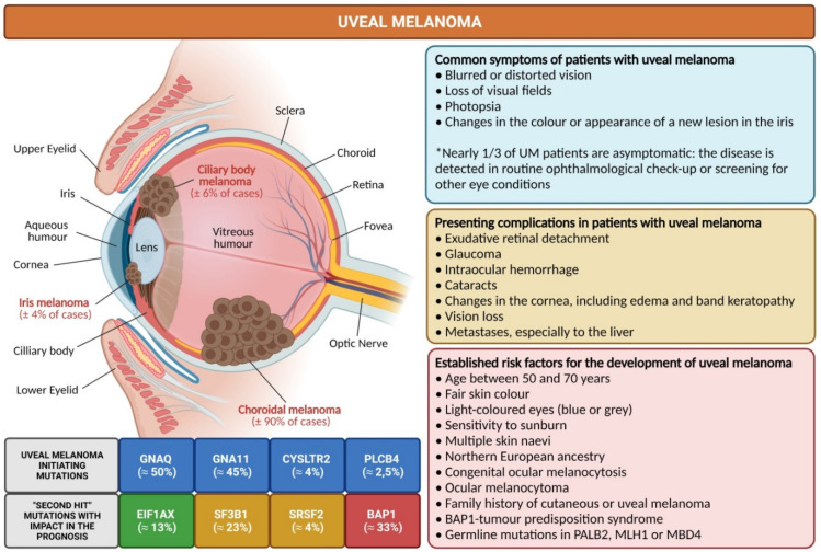
Uveal melanoma (UM) is the most common malignant intraocular tumour in the adult population. It is a rare cancer with an incidence of nearly five cases per million inhabitants per year, which develops from the uncontrolled proliferation of melanocytes in the choroid (≈90%), ciliary body (≈6%) or iris (≈4%). Patients initially present either with symptoms like blurred vision or photopsia, or without symptoms, with the tumour being detected in routine eye exams. Over the course of the disease, metastases, which are initially dormant, develop in nearly 50% of patients, preferentially in the liver. Despite decades of intensive research, the only approach proven to mildly control disease spread are early treatments directed to ablate liver metastases, such as surgical excision or chemoembolization. However, most patients have a limited life expectancy once metastases are detected, since there are limited therapeutic approaches for the metastatic disease, including immunotherapy, which unlike in cutaneous melanoma, has been mostly ineffective for UM patients. Therefore, in order to offer the best care possible to these patients, there is an urgent need to find robust models that can accurately predict the prognosis of UM, as well as therapeutic strategies that effectively block and/or limit the spread of the metastatic disease. Here, we initially summarized the current knowledge about UM by compiling the most relevant epidemiological, clinical, pathological and molecular data. Then, we revisited the most important prognostic factors currently used for the evaluation and follow-up of primary UM cases. Afterwards, we addressed emerging prognostic biomarkers in UM, by comprehensively reviewing gene signatures, immunohistochemistry-based markers and proteomic markers resulting from research studies conducted over the past three years. Finally, we discussed the current hurdles in the field and anticipated the future challenges and novel avenues of research in UM.
Keywords: biomarkers; metastases; molecular pathology; prognostic factors; survival; uveal melanoma.
Conflict of interest statement
The authors declare no conflict of interest.
Figures
Similar articles
-
Liver metastasis in uveal melanoma - treatment options and clinical outcome.Front Biosci (Landmark Ed). 2022 Feb 21;27(2):72. doi: 10.31083/j.fbl2702072. Front Biosci (Landmark Ed). 2022. PMID: 35227015 Review.
-
Molecular Insights and Emerging Strategies for Treatment of Metastatic Uveal Melanoma.Cancers (Basel). 2020 Sep 25;12(10):2761. doi: 10.3390/cancers12102761. Cancers (Basel). 2020. PMID: 32992823 Free PMC article. Review.
-
High Cysteinyl Leukotriene Receptor 1 Expression Correlates with Poor Survival of Uveal Melanoma Patients and Cognate Antagonist Drugs Modulate the Growth, Cancer Secretome, and Metabolism of Uveal Melanoma Cells.Cancers (Basel). 2020 Oct 13;12(10):2950. doi: 10.3390/cancers12102950. Cancers (Basel). 2020. PMID: 33066024 Free PMC article.
-
Prognostic impact of chromosomal aberrations and GNAQ, GNA11 and BAP1 mutations in uveal melanoma.Acta Ophthalmol. 2018 Feb;96(1):31-38. doi: 10.1111/aos.13452. Epub 2017 Apr 26. Acta Ophthalmol. 2018. PMID: 28444874
-
Limited value of 18F-FDG PET/CT and S-100B tumour marker in the detection of liver metastases from uveal melanoma compared to liver metastases from cutaneous melanoma.Eur J Nucl Med Mol Imaging. 2009 Nov;36(11):1774-82. doi: 10.1007/s00259-009-1175-0. Epub 2009 Jun 4. Eur J Nucl Med Mol Imaging. 2009. PMID: 19495748
Cited by
-
Uveal Melanoma Patients Have a Distinct Metabolic Phenotype in Peripheral Blood.Int J Mol Sci. 2023 Mar 7;24(6):5077. doi: 10.3390/ijms24065077. Int J Mol Sci. 2023. PMID: 36982149 Free PMC article.
-
Genome-Wide Methylation Patterns in Primary Uveal Melanoma: Development of MethylSig-UM, an Epigenomic Prognostic Signature to Improve Patient Stratification.Cancers (Basel). 2024 Jul 25;16(15):2650. doi: 10.3390/cancers16152650. Cancers (Basel). 2024. PMID: 39123378 Free PMC article.
-
Uveal Melanoma: Comprehensive Review of Its Pathophysiology, Diagnosis, Treatment, and Future Perspectives.Biomedicines. 2024 Aug 5;12(8):1758. doi: 10.3390/biomedicines12081758. Biomedicines. 2024. PMID: 39200222 Free PMC article. Review.
-
Assessment of ferroptosis as a promising candidate for metastatic uveal melanoma treatment and prognostication.Front Pharmacol. 2024 Oct 1;15:1466896. doi: 10.3389/fphar.2024.1466896. eCollection 2024. Front Pharmacol. 2024. PMID: 39411069 Free PMC article. Review.
-
Advances and Challenges in Immunotherapy for Metastatic Uveal Melanoma: Clinical Strategies and Emerging Targets.J Clin Med. 2025 Jul 19;14(14):5137. doi: 10.3390/jcm14145137. J Clin Med. 2025. PMID: 40725830 Free PMC article. Review.
References
Publication types
LinkOut - more resources
Full Text Sources
Miscellaneous
