. 2021 Dec 26;14(1):101.
doi: 10.3390/cancers14010101.

Automatic Segmentation of Metastatic Breast Cancer Lesions on 18F-FDG PET/CT Longitudinal Acquisitions for Treatment Response Assessment

Affiliations

Automatic Segmentation of Metastatic Breast Cancer Lesions on 18F-FDG PET/CT Longitudinal Acquisitions for Treatment Response Assessment

Noémie Moreau et al. Cancers (Basel). .

Abstract

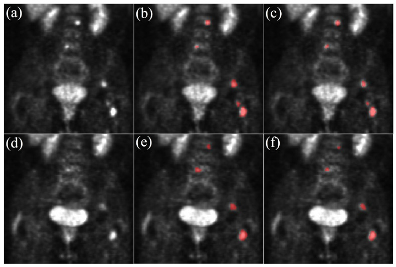
Metastatic breast cancer patients receive lifelong medication and are regularly monitored for disease progression. The aim of this work was to (1) propose networks to segment breast cancer metastatic lesions on longitudinal whole-body PET/CT and (2) extract imaging biomarkers from the segmentations and evaluate their potential to determine treatment response. Baseline and follow-up PET/CT images of 60 patients from the EPICUREseinmeta study were used to train two deep-learning models to segment breast cancer metastatic lesions: One for baseline images and one for follow-up images. From the automatic segmentations, four imaging biomarkers were computed and evaluated: SULpeak, Total Lesion Glycolysis (TLG), PET Bone Index (PBI) and PET Liver Index (PLI). The first network obtained a mean Dice score of 0.66 on baseline acquisitions. The second network obtained a mean Dice score of 0.58 on follow-up acquisitions. SULpeak, with a 32% decrease between baseline and follow-up, was the biomarker best able to assess patients' response (sensitivity 87%, specificity 87%), followed by TLG (43% decrease, sensitivity 73%, specificity 81%) and PBI (8% decrease, sensitivity 69%, specificity 69%). Our networks constitute promising tools for the automatic segmentation of lesions in patients with metastatic breast cancer allowing treatment response assessment with several biomarkers.

Keywords: automatic segmentation; deep learning; disease monitoring; imaging biomarkers; metastatic breast cancer.


Conflict of interest statement

Mario Campone received research grants via ICO institute from Pfizer, AstraZeneca, Sanofi, Gilead, Novartis, Lilly, Abbvie, Servier, Sandoz and Accord. All other authors certify that they have no affiliations with or involvement in any organization or entity with any financial or non-financial interest in the matter or materials discussed in this manuscript.

Figures

Figure 1
Figure 1
(a) U-NetBL and (b) U-NetFU networks’ architectures and inputs.
Figure 2
Figure 2
Segmentation examples on two acquisitions from the same patient. (a) PET BL, (b) GT BL, (c) U-NetBL, (d) PET FU, (e) GT FU, (f) U-NetFU. Zoom on the abdomen: kidneys, spine and bladder are visible. Due to the patient’s response to treatment, lesions on PET FU have a lower contrast than on PET BL and are less visible. BL = Baseline, GT = Ground Truth, FU = Follow-Up.
Figure 3
Figure 3
Segmentation examples on two acquisitions from 3 patients from the test dataset. (ac): Maximum intensity projections of PET images. (df): Ground truth segmentation overlaid on the maximum intensity projections of PET images. (gi): Automatic segmentation overlaid on the maximum intensity projections of PET images. U-NetBL was used on the baseline acquisition and U-NetFU on the follow-up acquisitions. For each pair of images: on the left the baseline acquisition and on the right the follow-up acquisition. DSC = dice score between the ground truth and the automatic segmentation. Blue arrows outline discrepancies between manual and automatic segmentations.
Figure 4
Figure 4
Graphical representation of each imaging biomarker with x axis biomarkers measured on ground truth segmentations and y axis biomarkers measured on automatic segmentations. The line represents perfect concordance. The concordance and the correlation are evaluated with the Lin’s concordance correlation coefficient (Lin’s CCC) and the Spearman’s rank correlation coefficient (Spearman cor) respectively.
Figure 5
Figure 5
Receiver Operating Characteristic (ROC) curve, responders (CR or PR) vs. non-responders (SD or PD).
Figure 6
Figure 6
Imaging biomarkers assessment for one patient with partial response. (a) Maximum intensity projection of three PET acquisitions with their biomarkers measured using the automatic segmentation. (b) Graphical representation of each biomarker evaluation across 3 acquisitions (in percentage of the biomarkers from the baseline). BL for Baseline and FU for Follow-up.

Similar articles

Cited by

References

    1. O’Shaughnessy J. Extending survival with chemotherapy in metastatic breast cancer. Oncologist. 2005;10:20–29. doi: 10.1634/theoncologist.10-90003-20. - DOI - PubMed
    1. Sundquist M., Brudin L., Tejler G. Improved survival in metastatic breast cancer 1985–2016. Breast. 2017;31:46–50. doi: 10.1016/j.breast.2016.10.005. - DOI - PubMed
    1. Eisenhauer E.A., Therasse P., Bogaerts J., Schwartz L.H., Sargent D., Ford R., Dancey J., Arbuck S., Gwyther S., Mooney M., et al. New response evaluation criteria in solid tumours: Revised RECIST guideline (version 1.1) Eur. J. Cancer. 2009;45:228–247. doi: 10.1016/j.ejca.2008.10.026. - DOI - PubMed
    1. Schwartz L.H., Litière S., De Vries E., Ford R., Gwyther S., Mandrekar S., Shankar L., Bogaerts J., Chen A., Dancey J., et al. RECIST 1.1—Upyear and clarification: From the RECIST committee. Eur. J. Cancer. 2016;62:132–137. doi: 10.1016/j.ejca.2016.03.081. - DOI - PMC - PubMed
    1. Yang H.L., Liu T., Wang X.M., Xu Y., Deng S.M. Diagnosis of bone metastases: A meta-analysis comparing 18-FDG PET, CT, MRI and bone scintigraphy. Eur. Radiol. 2011;21:2604–2617. doi: 10.1007/s00330-011-2221-4. - DOI - PubMed

LinkOut - more resources