Cancer-Associated Stromal Cells Promote the Contribution of MMP2-Positive Bone Marrow-Derived Cells to Oral Squamous Cell Carcinoma Invasion
- PMID: 35008304
- PMCID: PMC8750016
- DOI: 10.3390/cancers14010137
Cancer-Associated Stromal Cells Promote the Contribution of MMP2-Positive Bone Marrow-Derived Cells to Oral Squamous Cell Carcinoma Invasion
Abstract
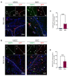
Tumor stromal components contribute to tumor development and invasion. However, the role of stromal cells in the contribution of bone marrow-derived cells (BMDCs) in oral squamous cell carcinoma (OSCC) invasion is unclear. In the present study, we created two different invasive OSCC patient-derived stroma xenografts (PDSXs) and analyzed and compared the effects of stromal cells on the relation of BMDCs and tumor invasion. We isolated stromal cells from two OSCC patients: less invasive verrucous OSCC (VSCC) and highly invasive conventional OSCC (SCC) and co-xenografted with the OSCC cell line (HSC-2) on green fluorescent protein (GFP)-positive bone marrow (BM) cells transplanted mice. We traced the GFP-positive BM cells by immunohistochemistry (IHC) and detected matrix metalloproteinase 2 (MMP2) expression on BM cells by double fluorescent IHC. The results indicated that the SCC-PDSX promotes MMP2-positive BMDCs recruitment to the invasive front line of the tumor. Furthermore, microarray analysis revealed that the expressions of interleukin 6; IL-6 mRNA and interleukin 1 beta; IL1B mRNA were higher in SCC stromal cells than in VSCC stromal cells. Thus, our study first reports that IL-6 and IL1B might be the potential stromal factors promoting the contribution of MMP2-positive BMDCs to OSCC invasion.
Keywords: MMP2; bone marrow-derived cells (BMDCs); oral squamous cell carcinoma invasion; patient-derived stromal cell xenograft (PDSX); stromal factor IL-6; stromal factor IL1B.
Conflict of interest statement
The authors declare no conflict of interest.
Figures


Similar articles
-
SOD3 Expression in Tumor Stroma Provides the Tumor Vessel Maturity in Oral Squamous Cell Carcinoma.Biomedicines. 2022 Oct 28;10(11):2729. doi: 10.3390/biomedicines10112729. Biomedicines. 2022. PMID: 36359248 Free PMC article.
-
Characterization and potential roles of bone marrow-derived stromal cells in cancer development and metastasis.Int J Med Sci. 2018 Sep 7;15(12):1406-1414. doi: 10.7150/ijms.24370. eCollection 2018. Int J Med Sci. 2018. PMID: 30275769 Free PMC article.
-
Stromal cells in the tumor microenvironment promote the progression of oral squamous cell carcinoma.Int J Oncol. 2021 Sep;59(3):72. doi: 10.3892/ijo.2021.5252. Epub 2021 Aug 9. Int J Oncol. 2021. PMID: 34368860 Free PMC article.
-
Differentiation and roles of bone marrow-derived cells on the tumor microenvironment of oral squamous cell carcinoma.Oncol Lett. 2019 Dec;18(6):6628-6638. doi: 10.3892/ol.2019.11045. Epub 2019 Nov 4. Oncol Lett. 2019. PMID: 31807176 Free PMC article.
-
Significance of cancer stroma for bone destruction in oral squamous cell carcinoma using different cancer stroma subtypes.Oncol Rep. 2022 Apr;47(4):81. doi: 10.3892/or.2022.8292. Epub 2022 Feb 25. Oncol Rep. 2022. PMID: 35211756 Free PMC article.
Cited by
-
Enzyme-Cleaved Bone Marrow Transplantation Improves the Engraftment of Bone Marrow Mesenchymal Stem Cells.JBMR Plus. 2023 Feb 11;7(3):e10722. doi: 10.1002/jbm4.10722. eCollection 2023 Mar. JBMR Plus. 2023. PMID: 36936364 Free PMC article.
-
SOD3 Expression in Tumor Stroma Provides the Tumor Vessel Maturity in Oral Squamous Cell Carcinoma.Biomedicines. 2022 Oct 28;10(11):2729. doi: 10.3390/biomedicines10112729. Biomedicines. 2022. PMID: 36359248 Free PMC article.
-
The Multiple Roles of CD147 in the Development and Progression of Oral Squamous Cell Carcinoma: An Overview.Int J Mol Sci. 2022 Jul 28;23(15):8336. doi: 10.3390/ijms23158336. Int J Mol Sci. 2022. PMID: 35955471 Free PMC article. Review.
References
-
- Sakuma K., Takahashi H., Kii T., Watanabe M., Tanaka A. Establishment and characterization of the human tongue squamous cell carcinoma cell line nokt-1. J. Hard. Tissue Biol. 2021;30:97–106. doi: 10.2485/jhtb.30.97. - DOI
-
- Meccariello G., Maniaci A., Bianchi G., Cammaroto G., Iannella G., Catalano A., Sgarzani R., De Vito A., Capaccio P., Pelucchi S., et al. Neck dissection and trans oral robotic surgery for oropharyngeal squamous cell carcinoma. Auris Nasus Larynx. 2021 doi: 10.1016/j.anl.2021.05.007. in press . - DOI - PubMed
Grants and funding
LinkOut - more resources
Full Text Sources
Research Materials
Miscellaneous

