. 2021 Dec 30;14(1):175.
doi: 10.3390/cancers14010175.

E-Cadherin-Deficient Epithelial Cells Are Sensitive to HDAC Inhibitors

Affiliations

E-Cadherin-Deficient Epithelial Cells Are Sensitive to HDAC Inhibitors

Lyvianne Decourtye-Espiard et al. Cancers (Basel). .

Abstract

Inactivating germline mutations in the CDH1 gene (encoding the E-cadherin protein) are the genetic hallmark of hereditary diffuse gastric cancer (HDGC), and somatic CDH1 mutations are an early event in the development of sporadic diffuse gastric cancer (DGC) and lobular breast cancer (LBC). In this study, histone deacetylase (HDAC) inhibitors were tested for their ability to preferentially inhibit the growth of human cell lines (MCF10A and NCI-N87) and murine organoids lacking CDH1 expression. CDH1-/- breast and gastric cells were more sensitive to the pan-HDAC inhibitors entinostat, pracinostat, mocetinostat and vorinostat than wild-type cells, with an elevated growth inhibition that was, in part, attributable to increased apoptosis. CDH1-null cells were also sensitive to more class-specific HDAC inhibitors, but compared to the pan-inhibitors, these effects were less robust to genetic background. Increased sensitivity to entinostat was also observed in gastric organoids with both Cdh1 and Tp53 deletions. However, the deletion of Tp53 largely abrogated the sensitivity of the Cdh1-null organoids to pracinostat and mocetinostat. Finally, entinostat enhanced Cdh1 expression in heterozygous Cdh1+/- murine organoids. In conclusion, entinostat is a promising drug for the chemoprevention and/or treatment of HDGC and may also be beneficial for the treatment of sporadic CDH1-deficient cancers.

Keywords: CDH1; E-cadherin; HDAC inhibitors; HDGC; synthetic lethality.


Conflict of interest statement

The authors declare no conflict of interest.

Figures

Figure 1
Figure 1
TCGA and STAD database highlight the correlation between HDACs and CDH1 expression. The relationship between HDAC classes and CDH1 expression from the Cancer Genome Atlas (TCGA) and the Stomach Adenocarcinoma (STAD) database was analyzed and the Spearman correlation was calculated between each HDAC and CDH1 (detailed values in Table S4). A low expression is represented in blue, and a high expression in red. (A) Hierarchical clustering of zinc and NAD+-dependent HDACs. Correlation between HDACs and CDH1 for (B) Class I HDAC (HDAC1, HDAC2, HDAC3 and HDAC8), (C) Class IIa HDAC (HDAC4, HDAC5, HDAC7 and HDAC9), (D) Class IIb (HDAC6 and HDAC10), (E) Class III HDAC (Sirtuins) and (F) Class IV HDAC (HDAC11). The HDACs with a significant correlation with CDH1 expression are labeled with an asterisk (*). LC = Lauren Classification, representing the different gastric cancer subtypes: red, diffuse; green, intestinal; and blue, mixed subtypes.
Figure 2
Figure 2
Pan-HDAC inhibitors have a synthetic lethal effect in gastric cancer cells lacking E-cadherin. A drugging experiment was performed on NCI-N87-WT and NCI-N87-CDH1−/− cells, and nuclei count was measured after 48 h. (A) Entinostat induced a synthetic lethal effect from 0.63 μM, with a maximum of 36% differences between both groups at 2.5 μM. (B) Effect of pracinostat was significant from 0.31 μM, with a maximum difference of 29%. (C) Mocetinostat was significant from 0.31 μM, with a maximum difference of 38%. (D) Vorinostat induced a synthetic lethal effect from 0.63 to 10 μM, with a maximum effect at 2.5 μM, with 23% differences between the two groups; n = 3–5 per compound. All the IC50 are summarized in Table S5. Statistically significant results are labeled as * p < 0.05, ** p < 0.01 and *** p < 0.001.
Figure 3
Figure 3
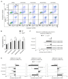
Pan-HDAC inhibitors preferentially induce apoptosis and decrease proliferation in gastric E-cadherin-deficient cells. Apoptosis, proliferation and cell cycle were analyzed in NCI-N87-CDH1−/− and NCI-N87-WT cells after 48 h of drugging with pan-HDAC inhibitors. (A) Apoptosis was analyzed by flow cytometry, using Annexin-V-FITC/propidium iodide staining. Annexin-V-FITC labels a membrane protein expressed by early apoptotic cells and propidium iodide stains nuclear DNA only accessible in apoptotic cells that have lost their membrane permeability (late apoptosis, n = 4–8). (B) Percentage of total apoptosis (early and late apoptosis combined) was increased in NCI-N87-CDH1−/− compared to NCI-N87-WT cells after 2.5 μM entinostat, 0.3 μM pracinostat and 2.5 μM vorinostat drugging (n = 3–5 per compound). (C) Total proliferation in NCI-N87-CDH1−/− and NCI-N87-WT cells was analyzed by flow cytometry after 48 h of drugging with pan-HDAC inhibitors. The difference in KI-67-positive cells for each drug was normalized compared to their respective DMSO. Mocetinostat (0.6 μM) and vorinostat (2.5 μM) preferentially and significantly decreased proliferation in NCI-N87-CDH1−/− cells compared to WT ones (n = 4–7); * = p-value < 0.05 for CDH1−/− vs. WT and ≠ from 0 for value statistically different to their respective DMSO (value of 0%). (D) Difference in percentage of cells in each phase of the cell cycle compared to their respective DMSO by flow cytometry, using propidium iodide staining (n = 4–5). Pracinostat and vorinostat significantly decreased the percentage of NCI-N87-CDH1−/− cells in G1 phase and rationally increased the proportion of cells in the S phase.
Figure 4
Figure 4
Cdh1 and Tp53 inactivation disrupt gastric organoids morphology. Organoids were generated using inducible Cre-lox mice. WT organoids express an inducible tdTomato fluorescence following Cre induction. Cdh1−/− organoids exhibit tdTomato expression and Cdh1 deletion while Cdh1−/−/Tp53−/− organoids exhibit tdTomato expression associated with Cdh1 and Tp53 deletion when induced with endoxifen. Organoids were induced at day 1 post-seeding followed by Western blot and immunofluorescences at day 6. (A) Western blot was performed on protein extracted from WT, Cdh1−/− and Cdh1−/−/Tp53−/− organoids. E-cadherin staining was observed at 110 kDa, Tp53 at 53 kDa and β-actin at 45 kDa. (B) Bright field and tdTomato expression were assessed after 5 days of induction with endoxifen. WT organoids maintained a circular morphology while both Cdh1−/− and Cdh1−/−/Tp53−/− organoids displayed a loss of circularity with the presence of evaginations. (C) Immunofluorescence on WT, Cdh1−/− and Cdh1−/−/Tp53−/− organoids with E-cadherin stained in green, tdTomato in red and DAPI in blue for nuclear staining. The absence of E-cadherin was confirmed in both Cdh1−/− and Cdh1−/−/Tp53−/− organoids. (D) Immunofluorescence with Tp53 staining in green. Tp53 was present in the WT and Cdh1−/− organoids and absent in the Cdh1−/−/Tp53−/− organoids. (E) KI-67 to label for proliferation was performed at day 6 post-seeding. KI-67 (in green) was absent in the WT organoids at day 6 indicating an arrest of proliferation whereas Cdh1−/− and Cdh1−/−/Tp53−/− organoids still presented cells in proliferation. (F) The intensity ratio between KI-67 and DAPI staining was measured every 10 microns on whole WT, Cdh1−/− and Cdh1−/−/Tp53−/− organoids by confocal microscopy. Statistically significant results are labeled as * p < 0.05 and *** p < 0.001.
Figure 5
Figure 5
E-cadherin-deficient gastric organoids are more sensitive to pan-HDAC inhibitors. WT, Cdh1−/− and Cdh1−/−/Tp53−/− organoids were drugged for 48 h with the different pan-HDAC inhibitors, and the area of the organoids was then measured and normalized to their respective vehicle controls. RFP fluorescence was used to detect tdTomato and determine the area of the organoids, using Cytation 5 imager (Biotek, Winooski, VT, USA). (A) Entinostat induced a synthetic lethal effect with a maximum of 44% differences between WT organoids and Cdh1−/− ones and 40% for the Cdh1−/−/Tp53−/− organoids. (B) Pracinostat induced an SL effect up to 40% and 20% for the Cdh1−/− and the Cdh1−/−/Tp53−/− organoids respectively. (C) Mocetinostat preferentially targeted E-cadherin-deficient cells with a maximum of 39% decrease in Cdh1−/− area compared to WT ones and had no effect for Cdh1−/−/Tp53−/− organoids. (D) Vorinostat did not present any significant synthetic lethal effect between the WT and the E-cadherin-deficient gastric organoids. n = 3–5 per compound. All the IC50 are summarized in Table S5. Statistically significant results are labeled as * p < 0.05, ** p < 0.01 and *** p < 0.001. (E) Picture representation of WT, Cdh1−/− and Cdh1−/−/Trp53−/− organoids after 7.5 μM entinostat, 0.25 μM pracinostat, 0.63 μM mocetinostat and 0.63 μM vorinostat.
Figure 6
Figure 6
E-cadherin-deficient mammary organoids are more sensitive to pan-HDAC inhibitors. WT and Cdh1−/− mammary organoids were generated from the same Cre-lox mice than the gastric organoids and drugged with the pan-HDAC inhibitors. The mammary organoids area were then assessed after 48 h of drugging and normalized to their respective DMSO. (A) Entinostat induced a synthetic lethal effect, with a maximum of 32% differences at 15 µM, between WT and Cdh1−/− organoids. (B) Pracinostat decreased Cdh1−/− growth up to 35% at 0.25 µM compared to the WT. (C) Mocetinostat induced an SL effect, with a maximum of 24% decrease at 10 µM, in the Cdh1−/− area compared to WT mammary organoids. (D) Vorinostat presented an SL effect at the lowest concentration, with a difference of 19% between the Cdh1−/− and WT organoids; n = 3–5 per compound. All the IC50 are summarized in Table S5. Statistically significant results are labeled as * p < 0.05 and ** p < 0.01.
Figure 7
Figure 7
Entinostat increases E-cadherin expression in Cdh1+/− organoids, leading to rounded morphology. Cdh1+/− organoids were induced at day 1 followed by drugging at day2 with entinostat or DMSO for 48 h. (A) Bright field and tdTomato expression were assessed at day 4 in WT, Cdh1+/− and Cdh1−/− organoids. Cdh1 decrease of expression was associated with a loss of circularity and the presence of evagination. (B) Immunofluorescence was performed on Cdh1+/− organoids following 48 h of treatment with entinostat or its respective DMSO concentration. Bright field and tdTomato showed a maintenance of a circular morphology following entinostat treatment. Immunofluorescence on Cdh1+/− organoids was performed with E-cadherin stained in green, tdTomato in red and DAPI in blue for nuclear staining. A decrease of E-cadherin was observed after induction in the Cdh1+/− DMSO-treated organoids, whereas entinostat treated Cdh1+/− organoids maintained a strong E-cadherin expression. (C) Circularity of the Cdh1+/− organoids was measured after DMSO or entinostat drugging, with a value of 1 representing a perfect circle (n = 43–97 organoids analyzed). (D) E-cadherin expression after entinostat drugging was measured by flow cytometry for WT and Cdh1+/− gastric organoids and normalized to their respective DMSO (n = 4). Entinostat treatment increased the expression of E-cadherin in the Cdh1+/− organoids. Statistically significant results are labeled as * p < 0.05 and *** p < 0.001.

Similar articles

Cited by

References

    1. Blair V.R., McLeod M., Carneiro F., Coit D.G., D’Addario J.L., van Dieren J.M., Harris K.L., Hoogerbrugge N., Oliveira C., van der Post R.S., et al. Hereditary diffuse gastric cancer: Updated clinical practice guidelines. Lancet Oncol. 2020;21:e386–e397. doi: 10.1016/S1470-2045(20)30219-9. - DOI - PMC - PubMed
    1. Grady W.M., Willis J., Guilford P.J., Dunbier A.K., Toro T.T., Lynch H., Wiesner G., Ferguson K., Eng C., Park J.G., et al. Methylation of the CDH1 promoter as the second genetic hit in hereditary diffuse gastric cancer. Nat. Genet. 2000;26:16–17. doi: 10.1038/79120. - DOI - PubMed
    1. Bhatt T., Rizvi A., Batta S.P., Kataria S., Jamora C. Signaling and mechanical roles of E-cadherin. Cell Commun. Adhes. 2013;20:189–199. doi: 10.3109/15419061.2013.854778. - DOI - PubMed
    1. Carneiro P., Figueiredo J., Bordeira-Carrico R., Fernandes M.S., Carvalho J., Oliveira C., Seruca R. Therapeutic targets associated to E-cadherin dysfunction in gastric cancer. Expert Opin. Ther. Targets. 2013;17:1187–1201. doi: 10.1517/14728222.2013.827174. - DOI - PubMed
    1. Liu J., Sun X., Qin S., Wang H., Du N., Li Y., Pang Y., Wang C., Xu C., Ren H. CDH1 promoter methylation correlates with decreased gene expression and poor prognosis in patients with breast cancer. Oncol. Lett. 2016;11:2635–2643. doi: 10.3892/ol.2016.4274. - DOI - PMC - PubMed

LinkOut - more resources