Review
. 2022 Jan 4;14(1):238.
doi: 10.3390/cancers14010238.

The Functional Role of Extracellular Matrix Proteins in Cancer

Affiliations
Review

The Functional Role of Extracellular Matrix Proteins in Cancer

Nadezhda V Popova et al. Cancers (Basel). .

Abstract

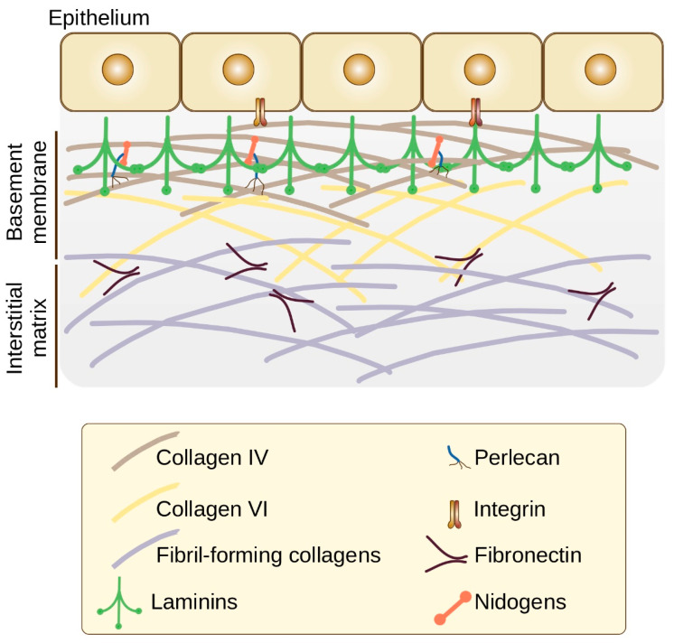
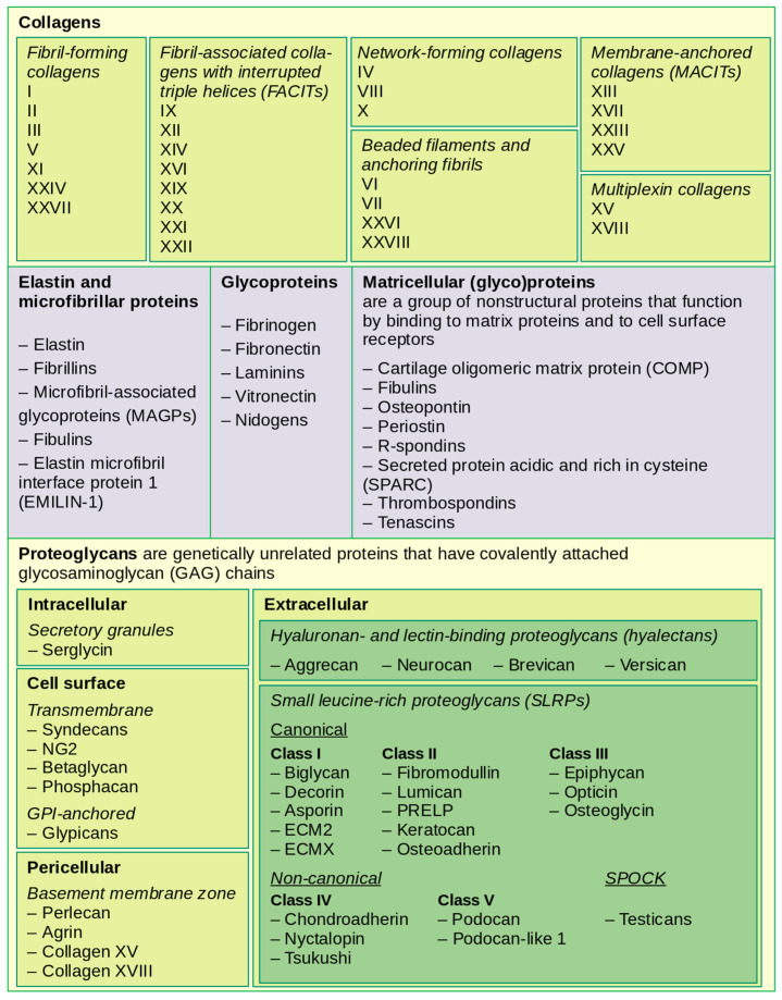
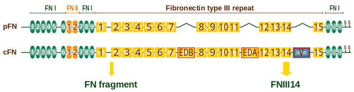
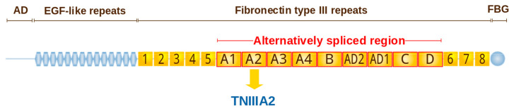
The extracellular matrix (ECM) is highly dynamic as it is constantly deposited, remodeled and degraded to maintain tissue homeostasis. ECM is a major structural component of the tumor microenvironment, and cancer development and progression require its extensive reorganization. Cancerized ECM is biochemically different in its composition and is stiffer compared to normal ECM. The abnormal ECM affects cancer progression by directly promoting cell proliferation, survival, migration and differentiation. The restructured extracellular matrix and its degradation fragments (matrikines) also modulate the signaling cascades mediated by the interaction with cell-surface receptors, deregulate the stromal cell behavior and lead to emergence of an oncogenic microenvironment. Here, we summarize the current state of understanding how the composition and structure of ECM changes during cancer progression. We also describe the functional role of key proteins, especially tenascin C and fibronectin, and signaling molecules involved in the formation of the tumor microenvironment, as well as the signaling pathways that they activate in cancer cells.

Keywords: collagen; extracellular matrix; fibronectin; matrikines; matrix metalloproteinases; tenascin; tumor microenvironment; tumor progression.


Conflict of interest statement

The authors declare no conflict of interest.

Figures

Figure 1
Figure 1
Schematic view of ECM organization (based on [12,17,18]). The major components of the basement membrane are the network-forming collagens (type IV collagen) and laminins. Nidogens and perlecan serve as binding bridges between the two networks: nidogens bridge laminin and collagen IV networks, and perlecan connects nidogen to collagen IV. Collagen VI interact with collagen IV, providing a link between the basement membrane and fibrillar components of the interstitial matrix.
Figure 2
Figure 2
Structural-functional classification of matrix proteins (based on [13,18,24,25,26]).
Figure 3
Figure 3
Domain structure of human fibronectin (based on SMART-tool [176] and review [177]). Each monomer contains three types of repeats: fibronectin type I (green ovals), type II (orange) and type III (gold rectangles). One or both of the FN type III modules (ED-A or ED-B) may be present in cellular fibronectin (cFN) but never in plasma fibronectin (pFN). A “variable” V or IIICS region is located between FNIII14 and FNIII15 and spliced out in ~50% subunits of pFN [178]. Two subunits are linked by a pair of C-terminal disulfide bonds to form a protein dimer. FNIII14 and “FN fragment” are proteolytic bioactive fragments derived from fibronectin.
Figure 4
Figure 4
Domain structure of human tenascin-C (based on SMART-tool [176] and review [226]). N-terminal assembly domain (AD) links the tenascin chains and mediates the oligomerization and formation of hexamers. The assembly domain is followed by the EGF-like repeats and two types of FN-III domains: conserved (yellow rectangles) and alternatively spliced (yellow with red), and the C-terminal fibrinogen-like domain (FBG). Tenascin-C-derived 22-mer peptide TNIIIA2.

Similar articles

Cited by

References

    1. Rowe R.G., Weiss S.J. Navigating ECM Barriers at the Invasive Front: The Cancer Cell-Stroma Interface. Annu. Rev. Cell Dev. Biol. 2009;25:567–595. doi: 10.1146/annurev.cellbio.24.110707.175315. - DOI - PubMed
    1. Piperigkou Z., Kyriakopoulou K., Koutsakis C., Mastronikolis S., Karamanos N.K. Key Matrix Remodeling Enzymes: Functions and Targeting in Cancer. Cancers. 2021;13:1441. doi: 10.3390/cancers13061441. - DOI - PMC - PubMed
    1. Ye M., Song Y., Pan S., Chu M., Wang Z.-W., Zhu X. Evolving Roles of Lysyl Oxidase Family in Tumorigenesis and Cancer Therapy. Pharmacol. Ther. 2020;215:107633. doi: 10.1016/j.pharmthera.2020.107633. - DOI - PubMed
    1. Dong Q., Liu X., Cheng K., Sheng J., Kong J., Liu T. Pre-Metastatic Niche Formation in Different Organs Induced by Tumor Extracellular Vesicles. Front. Cell Dev. Biol. 2021;9:733627. doi: 10.3389/fcell.2021.733627. - DOI - PMC - PubMed
    1. He C., Wang L., Li L., Zhu G. Extracellular Vesicle-Orchestrated Crosstalk between Cancer-Associated Fibroblasts and Tumors. Transl. Oncol. 2021;14:101231. doi: 10.1016/j.tranon.2021.101231. - DOI - PMC - PubMed

LinkOut - more resources