

Janus Kinase Inhibitors Ameliorated Gastrointestinal Amyloidosis and Hypoalbuminemia in Persistent Dermatitis Mouse Model
- PMID: 35008464
- PMCID: PMC8744922
- DOI: 10.3390/ijms23010028
Janus Kinase Inhibitors Ameliorated Gastrointestinal Amyloidosis and Hypoalbuminemia in Persistent Dermatitis Mouse Model
Abstract
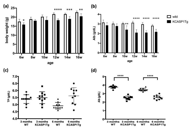
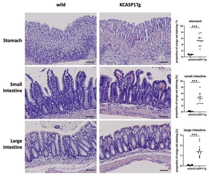
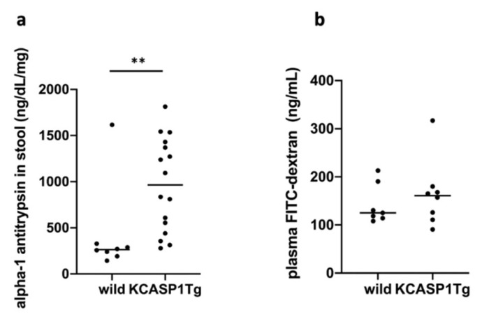
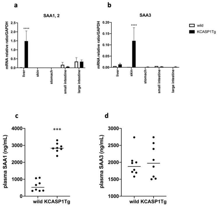
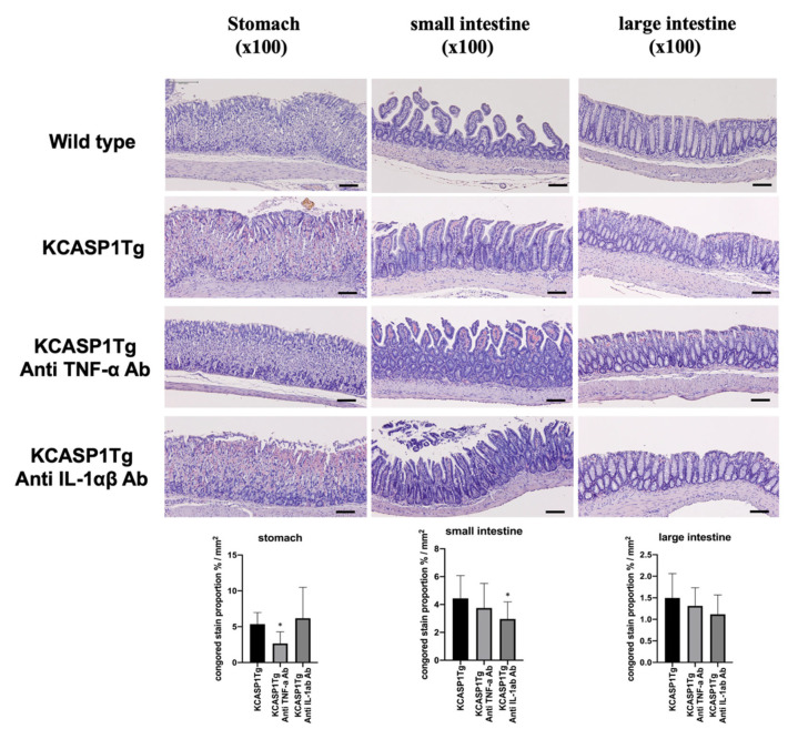
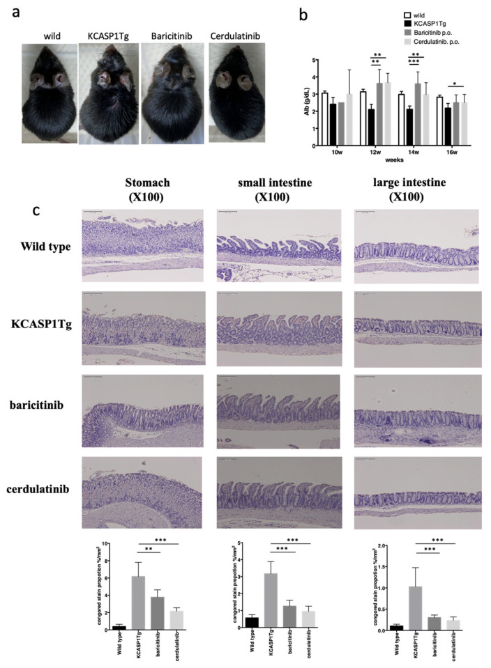
Malnutrition is not only regarded as a complication of rheumatoid arthritis and inflammatory bowel disease but also that of inflammatory skin disease; however, the mechanisms and efficacy of its treatment have not been elucidated. Using a mouse model of dermatitis, we investigated the pathophysiology of malnutrition in inflammatory skin conditions and efficacy of its treatment. We employed spontaneous skin inflammation mice models overexpressing human caspase-1 in the epidermal keratinocytes. Body weight, nutrition level, and α1-antitrypsin fecal concentration were measured. The gastrointestinal tract was histologically and functionally investigated. Fluorescein isothiocyanate (FITC)-dextran was forcibly fed on an empty stomach, and plasma FITC-dextran was measured. The treatment efficacy of antibodies against tumor necrosis factor-α (TNF-α) and interleukin (IL)-α/β as well as Janus kinase (JAK) inhibitors was investigated. Compared with wild-type littermates, the inflammatory skin mice models showed a lowered body weight, reduction of serum albumin level, amyloid deposition in the stomach, small intestine, and large intestine, and increased α1-antitrypsin fecal concentration. However, the plasma FITC-dextran was unchanged between the dermatitis models and wild-type littermates. The over-produced serum amyloid A1 in the liver was detected in the plasma in the dermatitis model. Antibodies against TNF-α and IL-α/β showed partial effects on amyloid deposition; however, JAK inhibitors improved gastrointestinal amyloidosis with the improvement of skin symptoms. Chronic dermatitis is closely related to secondary amyloidosis in the gastrointestinal tract, resulting in hypoalbuminemia. Therefore, active control of skin inflammation is essential for preventing gastrointestinal complications.
Keywords: JAK inhibitor; absorption; amyloidosis; cytokine; dermatitis; emaciation; gastro-intestinal tract; hypoalbuminemia; inflammatory skin mouse model; nutrition.
Conflict of interest statement
The authors declare no conflict of interest. The funders had no role in the design of the study; in the collection, analyses, or interpretation of data; in the writing of the manuscript, or in the decision to publish the results.
Figures
Similar articles
-
IL-17A Is the Critical Cytokine for Liver and Spleen Amyloidosis in Inflammatory Skin Disease.Int J Mol Sci. 2022 May 20;23(10):5726. doi: 10.3390/ijms23105726. Int J Mol Sci. 2022. PMID: 35628531 Free PMC article.
-
Amelioration of Systemic Amyloidosis by Blocking IL-17A and Not by IL-17F, and Arteriosclerosis by Blocking Both IL-17A and IL-17F in an Inflammatory Skin Mouse Model.Int J Mol Sci. 2024 Oct 29;25(21):11617. doi: 10.3390/ijms252111617. Int J Mol Sci. 2024. PMID: 39519167 Free PMC article.
-
Ruxolitinib Cream Has Dual Efficacy on Pruritus and Inflammation in Experimental Dermatitis.Front Immunol. 2021 Feb 15;11:620098. doi: 10.3389/fimmu.2020.620098. eCollection 2020. Front Immunol. 2021. PMID: 33658996 Free PMC article.
-
New JAK inhibitors for the treatment of psoriasis and psoriatic arthritis.G Ital Dermatol Venereol. 2020 Aug;155(4):411-420. doi: 10.23736/S0392-0488.20.06658-4. Epub 2020 Jun 15. G Ital Dermatol Venereol. 2020. PMID: 32545945
-
Systemic amyloidosis and the gastrointestinal tract.Nat Rev Gastroenterol Hepatol. 2009 Oct;6(10):608-17. doi: 10.1038/nrgastro.2009.147. Epub 2009 Sep 1. Nat Rev Gastroenterol Hepatol. 2009. PMID: 19724253 Review.
Cited by
-
IL-17A Is the Critical Cytokine for Liver and Spleen Amyloidosis in Inflammatory Skin Disease.Int J Mol Sci. 2022 May 20;23(10):5726. doi: 10.3390/ijms23105726. Int J Mol Sci. 2022. PMID: 35628531 Free PMC article.
-
Inflammatory Skin Disease Causes Anxiety Symptoms Leading to an Irreversible Course.Int J Mol Sci. 2023 Mar 21;24(6):5942. doi: 10.3390/ijms24065942. Int J Mol Sci. 2023. PMID: 36983014 Free PMC article.
-
Amelioration of Systemic Amyloidosis by Blocking IL-17A and Not by IL-17F, and Arteriosclerosis by Blocking Both IL-17A and IL-17F in an Inflammatory Skin Mouse Model.Int J Mol Sci. 2024 Oct 29;25(21):11617. doi: 10.3390/ijms252111617. Int J Mol Sci. 2024. PMID: 39519167 Free PMC article.
-
Arteriosclerosis Derived from Cutaneous Inflammation Is Ameliorated by the Deletion of IL-17A and IL-17F.Int J Mol Sci. 2023 Mar 12;24(6):5434. doi: 10.3390/ijms24065434. Int J Mol Sci. 2023. PMID: 36982506 Free PMC article.
-
Laggera alata Attenuates Inflammatory Response by Regulating Macrophage Polarization in Rheumatoid Arthritis Mice.Mol Biotechnol. 2024 Aug;66(8):1934-1941. doi: 10.1007/s12033-023-00808-w. Epub 2023 Jul 26. Mol Biotechnol. 2024. PMID: 37493934
References
-
- Couderc E., Morel F., Levillain P., Buffiere-Morgado A., Camus M., Paquier C., Bodet C., Jegou J.F., Pohin M., Favot L., et al. Interleukin-17A-induced production of acute serum amyloid A by keratinocytes contributes to psoriasis pathogenesis. PLoS ONE. 2017;12:e0181486. doi: 10.1371/journal.pone.0181486. - DOI - PMC - PubMed
-
- Yamanaka K., Nakanishi T., Saito H., Maruyama J., Isoda K., Yokochi A., Imanaka-Yoshida K., Tsuda K., Kakeda M., Okamoto R., et al. Persistent release of IL-1s from skin is associated with systemic cardio-vascular disease, emaciation and systemic amyloidosis: The potential of anti-IL-1 therapy for systemic inflammatory diseases. PLoS ONE. 2014;9:e104479. doi: 10.1371/journal.pone.0104479. - DOI - PMC - PubMed
-
- Kato S., Matsushima Y., Mizutani K., Kawakita F., Fujimoto M., Okada K., Kondo M., Habe K., Suzuki H., Mizutani H., et al. The Stenosis of Cerebral Arteries and Impaired Brain Glucose Uptake by Long-Lasting Inflammatory Cytokine Release from Dermatitis Is Rescued by Anti-IL-1 Therapy. J. Investig. Dermatol. 2018;138:2280–2283. doi: 10.1016/j.jid.2018.04.016. - DOI - PubMed
MeSH terms
Substances
Grants and funding
LinkOut - more resources
Full Text Sources
Medical
