

Cumulus Extracellular Matrix Is an Important Part of Oocyte Microenvironment in Ovarian Follicles: Its Remodeling and Proteolytic Degradation
- PMID: 35008478
- PMCID: PMC8744823
- DOI: 10.3390/ijms23010054
Cumulus Extracellular Matrix Is an Important Part of Oocyte Microenvironment in Ovarian Follicles: Its Remodeling and Proteolytic Degradation
Abstract
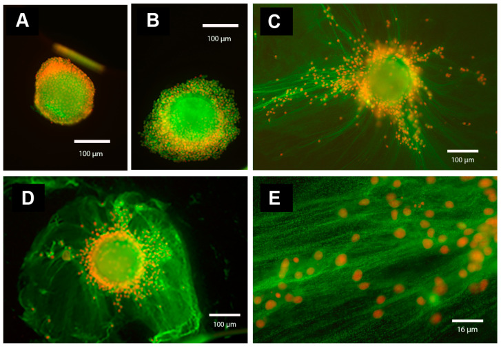
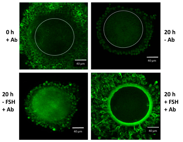
The extracellular matrix (ECM) is an essential structure with biological activities. It has been shown that the ECM influences gene expression via cytoskeletal components and the gene expression is dependent upon cell interactions with molecules and hormones. The development of ovarian follicles is a hormone dependent process. The surge in the luteinizing hormone triggers ovulatory changes in oocyte microenvironment. In this review, we discuss how proteolytic cleavage affects formation of cumulus ECM following hormonal stimulation; in particular, how the specific proteasome inhibitor MG132 affects gonadotropin-induced cytoskeletal structure, the organization of cumulus ECM, steroidogenesis, and nuclear maturation. We found that after the inhibition of proteolytic cleavage, gonadotropin-stimulated oocyte-cumulus complexes (OCCs) were without any signs of cumulus expansion; they remained compact with preserved cytoskeletal F-actin-rich transzonal projections through the oocyte investments. Concomitantly, a significant decrease was detected in progesterone secretion and in the expression of gonadotropin-stimulated cumulus expansion-related transcripts, such as HAS2 and TNFAIP6. In agreement, the covalent binding between hyaluronan and the heavy chains of serum-derived the inter-alpha-trypsin inhibitor, essential for the organization of cumulus ECM, was missing.
Keywords: extracellular matrix; hyaluronan; oocyte–cumulus complex; proteasome.
Conflict of interest statement
The authors declare no conflict of interest.
Figures
Similar articles
-
Inhibition of proteasomal proteolysis affects expression of extracellular matrix components and steroidogenesis in porcine oocyte-cumulus complexes.Domest Anim Endocrinol. 2012 Jan;42(1):50-62. doi: 10.1016/j.domaniend.2011.09.003. Epub 2011 Oct 8. Domest Anim Endocrinol. 2012. PMID: 22032857
-
Proteolytic activity of the 26S proteasome is required for the meiotic resumption, germinal vesicle breakdown, and cumulus expansion of porcine cumulus-oocyte complexes matured in vitro.Biol Reprod. 2008 Jan;78(1):115-26. doi: 10.1095/biolreprod.107.061366. Epub 2007 Oct 17. Biol Reprod. 2008. PMID: 17942798
-
The Biological Role of Hyaluronan-Rich Oocyte-Cumulus Extracellular Matrix in Female Reproduction.Int J Mol Sci. 2018 Jan 18;19(1):283. doi: 10.3390/ijms19010283. Int J Mol Sci. 2018. PMID: 29346283 Free PMC article. Review.
-
Organization of the expanded cumulus-extracellular matrix in preovulatory follicles: a role for inter-alpha-trypsin inhibitor.Endocr Regul. 2015 Jan;49(1):37-45. doi: 10.4149/endo_2015_01_37. Endocr Regul. 2015. PMID: 25687679 Review.
-
Unique hyaluronan structure of expanded oocyte-cumulus extracellular matrix in ovarian follicles.Endocr Regul. 2024 Aug 9;58(1):174-180. doi: 10.2478/enr-2024-0020. Print 2024 Jan 1. Endocr Regul. 2024. PMID: 39121477 Review.
Cited by
-
Effects of phthalate exposure on human ovarian extracellular matrix composition: insights from a 3D spheroid model.Environ Res. 2025 Aug 15;279(Pt 1):121797. doi: 10.1016/j.envres.2025.121797. Epub 2025 May 7. Environ Res. 2025. PMID: 40345415
-
Iron overload triggering ECM-mediated Hippo/YAP pathway in follicle development: a hypothetical model endowed with therapeutic implications.Front Endocrinol (Lausanne). 2023 May 8;14:1174817. doi: 10.3389/fendo.2023.1174817. eCollection 2023. Front Endocrinol (Lausanne). 2023. PMID: 37223010 Free PMC article. Review.
-
Ovarian stimulation with excessive FSH doses causes cumulus cell and oocyte dysfunction in small ovarian reserve heifers.Mol Hum Reprod. 2023 Sep 30;29(10):gaad033. doi: 10.1093/molehr/gaad033. Mol Hum Reprod. 2023. PMID: 37713463 Free PMC article.
-
Automatic Evaluation for Bioengineering of Human Artificial Ovary: A Model for Fertility Preservation for Prepubertal Female Patients with a Malignant Tumor.Int J Mol Sci. 2022 Oct 17;23(20):12419. doi: 10.3390/ijms232012419. Int J Mol Sci. 2022. PMID: 36293273 Free PMC article.
-
Dynamic Changes in Proteome during Yak Oocyte Maturation Analyzed Using iTRAQ Technology.Animals (Basel). 2023 Jun 23;13(13):2085. doi: 10.3390/ani13132085. Animals (Basel). 2023. PMID: 37443883 Free PMC article.
References
Publication types
MeSH terms
LinkOut - more resources
Full Text Sources
