. 2021 Dec 22;23(1):71.
doi: 10.3390/ijms23010071.

Elevation of Hyaluronan Synthase by Magnesium Supplementation Mediated through the Activation of GSK3 and CREB in Human Keratinocyte-Derived HaCaT Cells

Affiliations

Elevation of Hyaluronan Synthase by Magnesium Supplementation Mediated through the Activation of GSK3 and CREB in Human Keratinocyte-Derived HaCaT Cells

Kana Marunaka et al. Int J Mol Sci. .

Abstract

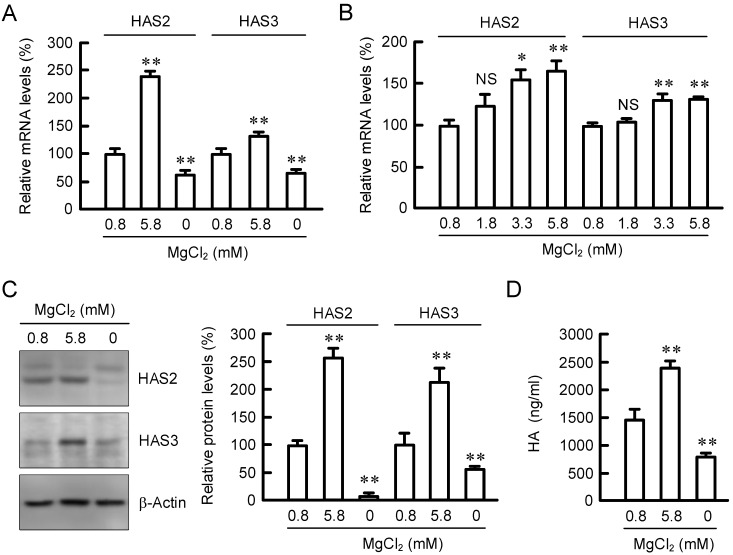
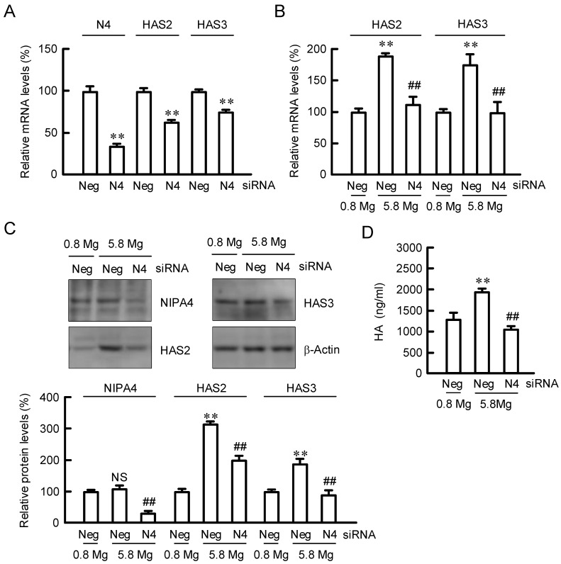
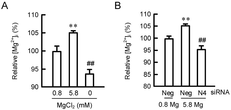
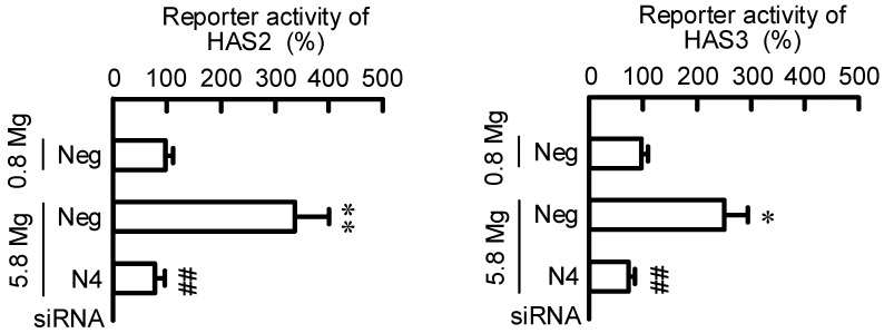
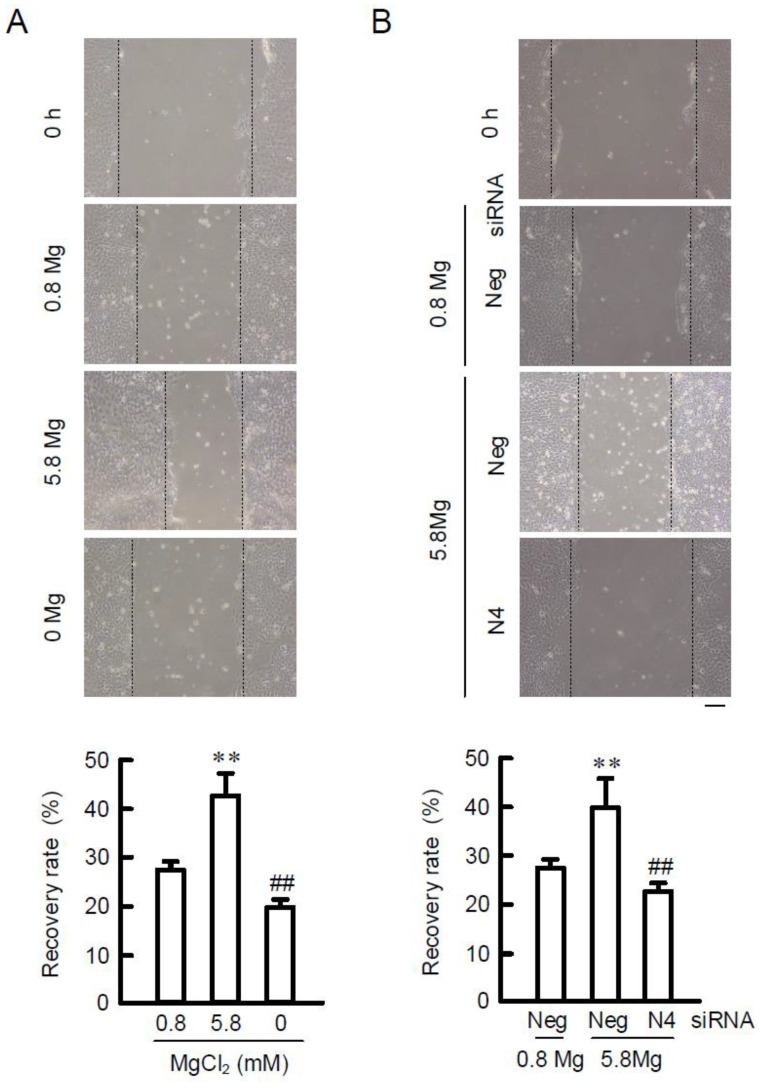
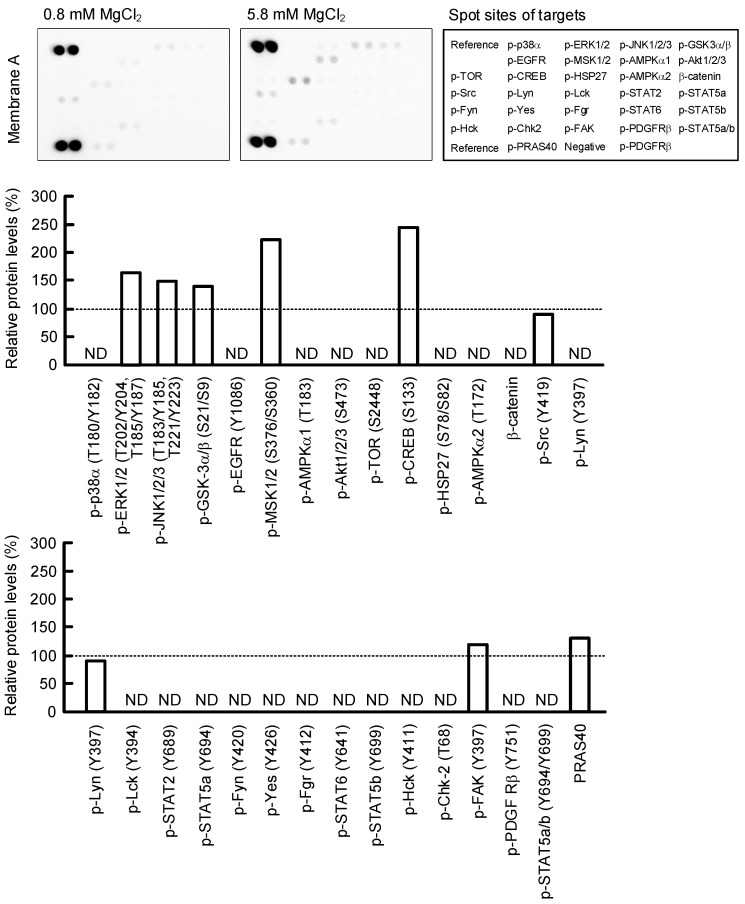
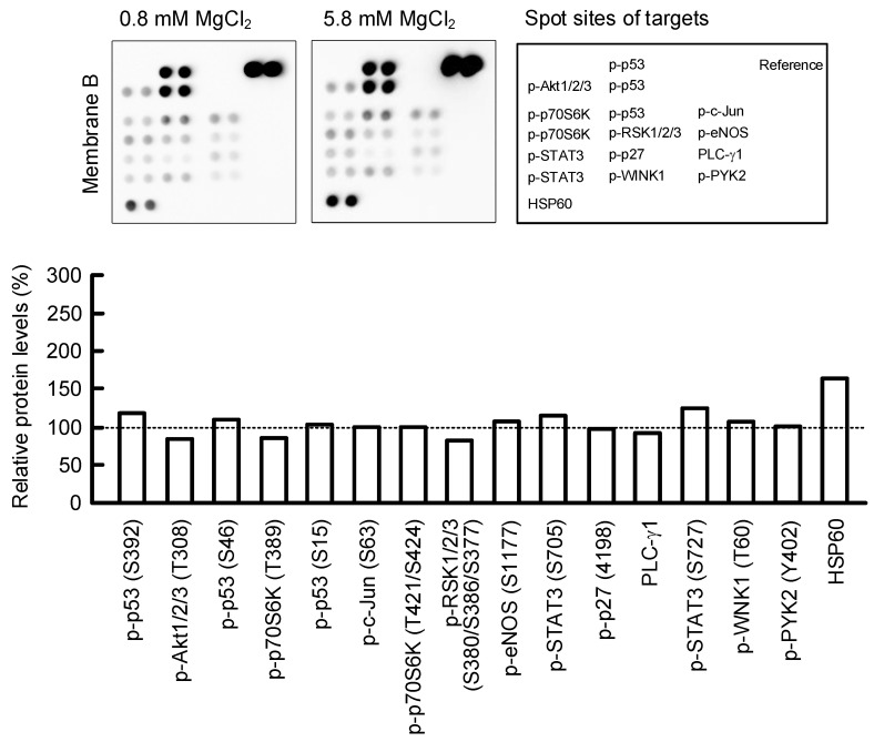
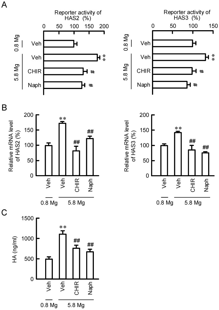
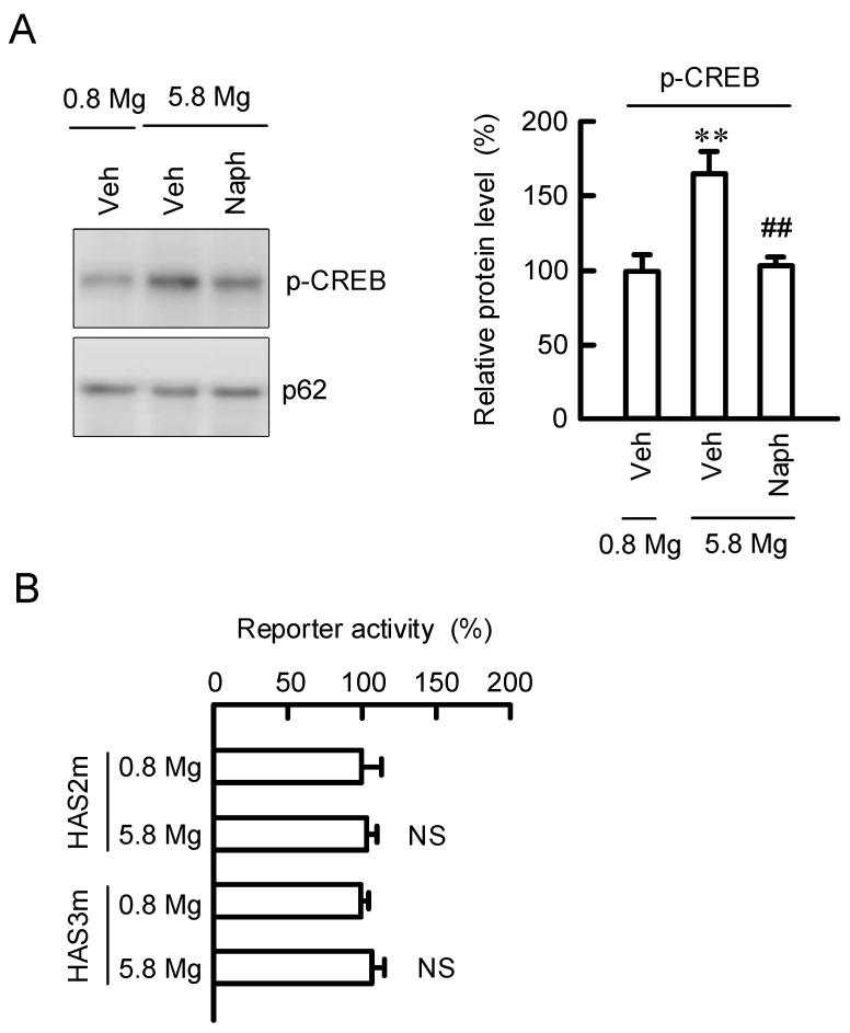
Skin barrier damage is present in the patients with hereditary disorders of the magnesium channel, but the molecular mechanism has not been fully understood. We found that the expressions of hyaluronan synthase (HAS), HAS2 and HAS3 are influenced by MgCl2 concentration in human keratinocyte-derived HaCaT cells. The exposure of cells to a high concentration (5.8 mM) of MgCl2 induced the elevation of HAS2/3 expression, which was inhibited by mRNA knockdown of nonimprinted in Prader-Willi/Angelman syndrome-like domain containing 4 (NIPAL4). Similarly, the content of hyaluronic acid (HA) was changed according to MgCl2 concentration and the expression of NIPAL4. The MgCl2 supplementation increased the reporter activities of HAS2/3, which were inhibited by NIPAL4 knockdown, indicating that the expressions of HAS2/3 are up-regulated at the transcriptional level. The reporter activities and mRNA levels of HAS2/3, and the production of HA were inhibited by CHIR-99021, a glycogen synthase kinase-3 (GSK3) inhibitor, and naphthol AS-E, a cyclic AMP-response element binding protein (CREB) inhibitor. Furthermore, the mutation in putative CREB-binding sites of promoter region in HAS2/3 genes inhibited the MgCl2 supplementation-induced elevation of promoter activity. Our results indicate that the expressions of HAS2/3 are up-regulated by MgCl2 supplementation in HaCaT cells mediated through the activation of GSK3 and CREB. Magnesium may play a pivotal role in maintaining the skin barrier function and magnesium supplementation may be useful to enhance moisturization and wound repair in the skin.

Keywords: hyaluronan synthase; hyaluronic acid; magnesium.


Conflict of interest statement

The authors declare no conflict of interest.

Figures

Figure 1
Figure 1
Effects of extracellular Mg2+ concentration on HAS2/3 expressions in HaCaT cells. (A,B) Cells were incubated in the presence of 0, 0.8, and 5.8 mM MgCl2 for 6 h. The mRNA levels of HAS2/3 were measured by real-time PCR analysis and represented as a percentage of 0.8 mM MgCl2. (C,D) Cells were incubated in the presence of 0, 0.8, and 5.8 mM MgCl2 for 24 h. The protein levels of HAS2/3 were measured by Western blotting analysis and represented as a percentage of 0.8 mM MgCl2. HA contents in the media were measured using a Hyaluronan Quantification kit. n = 3–4. ** p < 0.01 and * p < 0.05 significantly different from 0.8 mM MgCl2. NS p > 0.05.
Figure 2
Figure 2
Effect of NIPAL4 siRNA on HAS2/3 expressions. Cells were transfected with negative (Neg) or NIAPL4 (N4) siRNA. (A) After transfection, the cells were incubated in the presence of 0.8 mM MgCl2 for 72 h. The mRNA levels of N4 and HAS2/3 were measured by real-time PCR analysis and represented as a percentage of negative siRNA. (B) After 66 h of transfection, the cells were incubated in the presence of 0.8 and 5.8 mM MgCl2 for 6 h. The mRNA levels of HAS2/3 were measured by real-time PCR analysis and represented as a percentage of 0.8 mM MgCl2. (C,D) After 48 h of transfection, the cells were incubated in the presence of 0.8 and 5.8 mM MgCl2 for 24 h. The protein levels of NIPA4 and HAS2/3 were measured by Western blotting analysis and represented as a percentage of 0.8 mM MgCl2. HA contents in the media were measured using a Hyaluronan Quantification kit. n = 3–4. ** p < 0.01 significantly different from negative siRNA or 0.8 mM MgCl2. NS p > 0.05. ## p < 0.01 significantly different from negative siRNA plus 5.8 mM MgCl2.
Figure 3
Figure 3
Effects of extracellular Mg2+ concentration and NIPAL4 siRNA on [Mg2+]i. (A) Cells were incubated in the presence of 0, 0.8, and 5.8 mM MgCl2 for 24 h. The cells were loaded with KMG-20-AM for 30 min at 37 °C. The fluorescence intensity of KMG-20 was measured using a plate reader. [Mg2+]i is represented as a percentage of 0.8 mM MgCl2. (B) Cells were transfected with negative (Neg) or NIPAL4 (N4) siRNA. After 48 h of transfection, the cells were incubated in the presence of 0.8 and 5.8 mM MgCl2 for 24 h. [Mg2+]i is represented as a percentage of 0.8 mM MgCl2. n = 4–6. ** p < 0.01 significantly different from 0.8 mM MgCl2. ## p < 0.01 significantly different from negative siRNA plus 5.8 mM MgCl2.
Figure 4
Figure 4
Effects of extracellular Mg2+ concentration and NIPAL4 siRNA on reporter activities of HAS2/3. Cells were transfected with negative (Neg) or NIAPL4 (N4) siRNA. After 66 h of transfection, the cells were incubated in the presence of 0.8 and 5.8 mM MgCl2 for 6 h. The activities of secreted luciferase and SEAP were measured using a Ready-To-Glow Dual Secreted Reporter Assay kit. The reporter activity is represented as a percentage of 0.8 mM MgCl2. n = 4. ** p < 0.01 and * p < 0.05 significantly different from 0.8 mM MgCl2. ## p < 0.01 significantly different from negative siRNA plus 5.8 mM MgCl2.
Figure 5
Figure 5
Effects of extracellular Mg2+ concentration and NIPAL4 siRNA on cell migration. (A) After reaching confluent densities, the cells were incubated in 0, 0.8, and 5.8 mM MgCl2 media for 24 h. Then, the cells were scratched with tip of a 200-μL pipette tip. The image of 0 h was taken at just after scratch. Other images were taken after 24 h. (B) Cells were transfected with negative (Neg) or NIPAL4 (N4) siRNA. After 66 h of transfection, the cells were incubated in 0.8 and 5.8 mM MgCl2 media for 24 h. Then, the cells were scratched with tip of a 200-μL pipette tip. The images were taken after 24 h. The recovery rate is presented as the percentage of scratch closure. Scale bar indicates 50 μm. n = 4–6. ** p < 0.01 significantly different from 0.8 mM MgCl2. ## p < 0.01 significantly different from 5.8 mM MgCl2 or negative siRNA plus 5.8 mM MgCl2.
Figure 6
Figure 6
Effect of MgCl2 supplementation on phosphorylation of intracellular signaling proteins. Cells were incubated in 0.8 and 5.8 mM MgCl2 media for 6 h. After collecting whole cell lysates, the aliquots were applied on a Proteome Profiler Human Phospho-Kinase Array Kit. Upper images indicate the membrane A and B, and spot sites of targets. Lower images indicate the phosphorylation level or total amount are represented as a percentage of 0.8 mM MgCl2. A signal below the detection limit is indicated by non-detectable (ND).
Figure 6
Figure 6
Effect of MgCl2 supplementation on phosphorylation of intracellular signaling proteins. Cells were incubated in 0.8 and 5.8 mM MgCl2 media for 6 h. After collecting whole cell lysates, the aliquots were applied on a Proteome Profiler Human Phospho-Kinase Array Kit. Upper images indicate the membrane A and B, and spot sites of targets. Lower images indicate the phosphorylation level or total amount are represented as a percentage of 0.8 mM MgCl2. A signal below the detection limit is indicated by non-detectable (ND).
Figure 7
Figure 7
Effects of MgCl2 supplementation and signaling inhibitors on HAS2/3 expressions and HA production. (A) Cells transfected with promoter reporter vectors for HAS2/3 were incubated in the absence (Veh) and presence of 10 μM CHIR-99021 (CHIR) or 10 μM naphthol AS-E (Naph) for 6 h. The reporter activities of HAS2/3 are represented as a percentage of 0.8 mM MgCl2. (B) Cells were incubated in the absence (Veh) and presence of 0.8 mM MgCl2, 5.8 mM MgCl2, 10 μM CHIR-99021 (CHIR) or 10 μM naphthol AS-E (Naph) for 6 h. The mRNA levels of HAS2/3 are represented as a percentage of 0.8 mM MgCl2. (C) Cells were incubated in the absence (Veh) and presence of 0.8 mM MgCl2, 5.8 mM MgCl2, 10 μM CHIR-99021 (CHIR) or 10 μM naphthol AS-E (Naph) for 24 h. After collecting the media, HA contents were measured using a Hyaluronan Quantification kit. n = 4–6. ** p < 0.01 significantly different from 0.8 mM MgCl2. ## p < 0.01 and # p < 0.05 significantly different from 5.8 mM MgCl2 plus 5.8 mM MgCl2.
Figure 8
Figure 8
Involvement of CREB in the elevation of HAS2/3 expressions by MgCl2 supplementation. (A) Cells were incubated in the absence (Veh) and presence of 0.8 mM MgCl2, 5.8 mM MgCl2, or 10 μM naphthol AS-E (Naph) for 2 h. After isolation of nuclear fraction, the protein levels of p-CREB and nucleoporin p62 (p62), an internal control, were measured by Western blotting analysis and represented as a percentage of 0.8 mM MgCl2. (B) Cells were transfected with reporter vectors of HAS2/3 containing the mutation in CREB-binding site (HAS2m and HAS3m). Then, the cells were incubated in the presence of 0.8 and 5.8 mM MgCl2 for 6 h. The reporter activity is represented as a percentage of 0.8 mM MgCl2. n = 3–4. ** p < 0.01 significantly different from 0.8 mM MgCl2. NS p > 0.05. ## p < 0.01 significantly different from 5.8 mM MgCl2 plus 5.8 mM MgCl2.

References

    1. Oji V., Tadini G., Akiyama M., Bardon C.B., Bodemer C., Bourrat E., Coudiere P., DiGiovanna J.J., Elias P., Fischer J., et al. Revised nomenclature and classification of inherited ichthyoses: Results of the First Ichthyosis Consensus Conference in Soreze 2009. J. Am. Acad. Dermatol. 2010;63:607–641. doi: 10.1016/j.jaad.2009.11.020. - DOI - PubMed
    1. Wajid M., Kurban M., Shimomura Y., Christiano A.M. NIPAL4/ichthyin is expressed in the granular layer of human epidermis and mutated in two Pakistani families with autosomal recessive ichthyosis. Dermatology. 2010;220:8–14. doi: 10.1159/000265757. - DOI - PMC - PubMed
    1. Honda Y., Kitamura T., Naganuma T., Abe T., Ohno Y., Sassa T., Kihara A. Decreased Skin Barrier Lipid Acylceramide and Differentiation-Dependent Gene Expression in Ichthyosis Gene Nipal4-Knockout Mice. J. Investig. Dermatol. 2018;138:741–749. doi: 10.1016/j.jid.2017.11.008. - DOI - PubMed
    1. Pilchova I., Klacanova K., Tatarkova Z., Kaplan P., Racay P. The Involvement of Mg2+ in Regulation of Cellular and Mitochondrial Functions. Oxid. Med. Cell. Longev. 2017;2017:6797460. - PMC - PubMed
    1. Dabrowska A.K., Spano F., Derler S., Adlhart C., Spencer N.D., Rossi R.M. The relationship between skin function, barrier properties, and body-dependent factors. Skin Res. Technol. 2018;24:165–174. doi: 10.1111/srt.12424. - DOI - PubMed