. 2021 Dec 23;23(1):148.
doi: 10.3390/ijms23010148.

Exercise Preconditioning Blunts Early Atrogenes Expression and Atrophy in Gastrocnemius Muscle of Hindlimb Unloaded Mice

Affiliations

Exercise Preconditioning Blunts Early Atrogenes Expression and Atrophy in Gastrocnemius Muscle of Hindlimb Unloaded Mice

Lorenza Brocca et al. Int J Mol Sci. .

Abstract

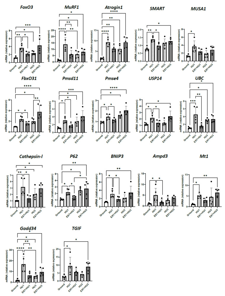
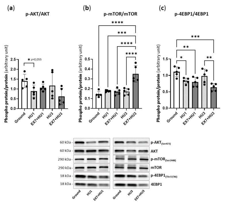
A large set of FoxOs-dependent genes play a primary role in controlling muscle mass during hindlimb unloading. Mitochondrial dysfunction can modulate such a process. We hypothesized that endurance exercise before disuse can protect against disuse-induced muscle atrophy by enhancing peroxisome proliferator-activated receptor-γ coactivator-1α (PGC1α) expression and preventing mitochondrial dysfunction and energy-sensing AMP-activated protein kinase (AMPK) activation. We studied cross sectional area (CSA) of muscle fibers of gastrocnemius muscle by histochemistry following 1, 3, 7, and 14 days of hindlimb unloading (HU). We used Western blotting and qRT-PCR to study mitochondrial dynamics and FoxOs-dependent atrogenes' expression at 1 and 3 days after HU. Preconditioned animals were submitted to moderate treadmill exercise for 7 days before disuse. Exercise preconditioning protected the gastrocnemius from disuse atrophy until 7 days of HU. It blunted alterations in mitochondrial dynamics up to 3 days after HU and the expression of most atrogenes at 1 day after disuse. In preconditioned mice, the activation of atrogenes resumed 3 days after HU when mitochondrial dynamics, assessed by profusion and pro-fission markers (mitofusin 1, MFN1, mitofusin 2, MFN2, optic atrophy 1, OPA1, dynamin related protein 1, DRP1 and fission 1, FIS1), PGC1α levels, and AMPK activation were at a basal level. Therefore, the normalization of mitochondrial dynamics and function was not sufficient to prevent atrogenes activation just a few days after HU. The time course of sirtuin 1 (SIRT1) expression and content paralleled the time course of atrogenes' expression. In conclusion, seven days of endurance exercise counteracted alterations of mitochondrial dynamics and the activation of atrogenes early into disuse. Despite the normalization of mitochondrial dynamics, the effect on atrogenes' suppression died away within 3 days of HU. Interestingly, muscle protection lasted until 7 days of HU. A longer or more intense exercise preconditioning may prolong atrogenes suppression and muscle protection.

Keywords: atrogenes; disuse atrophy; hindlimb unloading; physical preconditioning.


Conflict of interest statement

The authors declare no conflict of interest.

Figures

Figure 1
Figure 1
A single exercise bout and 7-day endurance training increase mRNA and protein expression of PGC 1α in ground mice. Gene expression and protein content analysis of PGC 1α (a) and activation level of AMP-kinase (b) in gastrocnemius of mice subjected to a single bout of treadmill exercise and relative representative Western blot; protein levels were normalized for tubulin expression. Ground: Control mice; Ground SB-EX + 0 h: Mice subjected to a single bout of exercise sacrificed immediately or 3 h after exercise (Ground SB-EX + 3 h). Gene expression and protein content analysis of PGC 1α (c) and activation level of AMP-kinase (d) in gastrocnemius of trained mice and relative representative Western blot; protein levels were normalized for tubulin expression. Ground: Control mice; Ground EX7 + 3 h: Mice subjected to 7 days of endurance exercise sacrificed 3 h after the last section of exercise. The activation level of AMP-kinase was determined through the ratio between the content of the phosphorylated (p) and total form. Data are presented as means ± SD * p < 0.05 *** p < 0.0005 **** p < 0.0001.
Figure 2
Figure 2
Physical preconditioning mitigates disuse-induced atrophy of gastrocnemius. Cross Sectional Area (CSA) of gastrocnemius muscle fibers of mice subjected to disuse alone and preceded by physical preconditioning and representative hematoxylin–eosin staining; scale bars: 100µm (a). CSA time course changes with disuse alone and preceded by 7 days of physical preconditioning (b). Ground: Control mice; HU1, HU3, HU7, and HU14: Mice subjected to 1, 3, 7, and 14 days of hindlimb unloading; Ground EX7: Mice subjected to 7 days of physical exercise. Data are presented as means ± SD * p < 0.05 ** p < 0.005 *** p < 0.0005. §: Different from ground and unexercised HU1 groups; †: Different from preconditioned HU3 group; ∗: Different from ground and preconditioned HU1 and HU3 groups.
Figure 3
Figure 3
Physical preconditioning counteracts the early atrogenes’ induction during disuse. RT-PCR analysis of transcriptional levels of FoxO3 and the muscle-specific ubiquitin ligases MuRF1, Atrogin1, SMART, MUSA1, FbxO31, proteasome subunit atrogenes (Psmd11, Psme4), de-ubiquitinating enzyme atrogene (USP14), ubiquitin atrogene (UBC), autophagy-related atrogenes (Cathepsin l, P62, BNIP3), AMP deaminase atrogene (Ampd3), oxidative stress related atrogene (Mt1), unfolded protein response atrogene (Gadd34), transcription regulators Smad2/3 atrogene (TGIF). The expression levels were normalized on GAPDH. Ground: Control mice; HU1: Mice subjected to 1 day of hindlimb unloading; EX7 + HU1: Mice subjected to 7 days of physical preconditioning followed by 1 day of hindlimb unloading; HU3: Mice subjected to 3 days of hindlimb unloading; EX7 + HU3: Mice subjected to 7 days of physical preconditioning followed by 3 days of hindlimb unloading. Data are presented as means ± SD * p < 0.05 ** p < 0.005 *** p < 0.0005 **** p < 0.0001.
Figure 4
Figure 4
Physical preconditioning effect on phosphorylation of kinases of the Akt/mTOR pathway during disuse. Determination of activity levels of p-AKT (a), p-mTOR (b), and p-4EBP1 (c) by Western blot analysis of the ratio between the content in the phosphorylated (p) and total forms and relative representative Western blot. Ground: Control mice; HU1: Mice subjected to 1 day of hindlimb unloading; EX7 + HU1: Mice subjected to 7 days of physical preconditioning followed by 1 day of hindlimb unloading; HU3: Mice subjected to 3 days of hindlimb unloading; EX7 + HU3: Mice subjected to 7 days of physical preconditioning followed by 3 days of hindlimb unloading. Data are presented as means ± SD * p < 0.05 ** p < 0.005 *** p < 0.0005 **** p < 0.0001.
Figure 5
Figure 5
Physical preconditioning counteracts the decrease in MFN2 pro-fusion protein induced by disuse. Quantification of protein content of mitochondria dynamics markers by Western blotting. Pro-fusion proteins MFN1 (a), MFN2 (b), OPA1 (c), pro-fission proteins p-DRP1(Ser616) (d), p-DRP1(Ser637) (e), and FIS1 (f), and relative representative Western blot. Ground: Control mice; HU1: Mice subjected to 1 day of hindlimb unloading; EX7 + HU1: Mice subjected to 7 days of physical preconditioning followed by 1 day of hindlimb unloading; HU3: Mice subjected to 3 days of hindlimb unloading; EX7 + HU3: Mice subjected to 7 days of physical preconditioning followed by 3 days of hindlimb unloading. Protein levels were normalized for tubulin expression. The phosphorylation status of DRP1(Ser616) and DRP1(Ser637) was determined through the ratio between the content in the phosphorylated and total forms. Data are presented as means ± SD * p < 0.05 ** p < 0.005 *** p < 0.0005.
Figure 6
Figure 6
Effects of hindlimb unloading and exercise preconditioning on FoxO regulators. Activation level of AMP-kinase (a), Gene expression and protein content analysis of PGC1α (b), gene expression and protein content analysis of SIRT1 (c), and relative representative Western blot. Ground: Control mice; HU1: Mice subjected to 1 day of hindlimb unloading; EX7 + HU1: Mice subjected to 7 days of physical preconditioning followed by 1 day of hindlimb unloading; HU3: Mice subjected to 3 days of hindlimb unloading; EX7 + HU3: Mice subjected to 7 days of physical preconditioning followed by 3 days of hindlimb unloading. Data are presented as means ± SD * p < 0.05 ** p < 0.005 *** p < 0.0005 **** p < 0.0001. Gene expression levels were determined by RT-PCR analysis and normalized on GAPDH. Protein content levels were determined by Western blot analysis and normalized for tubulin. The activation level of AMP-kinase was determined by Western blot analysis of the ratio between the content of the phosphorylated and total form.

References

    1. Mazzucco S., Agostini F., Biolo G. Inactivity-mediated insulin resistance is associated with upregulated pro-inflammatory fatty acids in human cell membranes. Clin. Nutr. 2010;29:386–390. doi: 10.1016/j.clnu.2009.09.006. - DOI - PubMed
    1. Booth F.W., Roberts C.K., Laye M.J. Lack of exercise is a major cause of chronic diseases. Compr. Physiol. 2012;2:1143–1211. - PMC - PubMed
    1. Bonaldo P., Sandri M. Cellular and molecular mechanisms of muscle atrophy. Dis. Model. Mech. 2013;6:25–39. doi: 10.1242/dmm.010389. - DOI - PMC - PubMed
    1. Pedersen B.K., Saltin B. Exercise as medicine-evidence for prescribing exercise as therapy in 26 different chronic diseases. Scand. J. Med. Sci. Sports. 2015;25((Suppl. 3)):1–72. doi: 10.1111/sms.12581. - DOI - PubMed
    1. Sandri M., Lin J., Handschin C., Yang W., Arany Z.P., Lecker S.H., Goldberg A.L., Spiegelman B.M. PGC-1alpha protects skeletal muscle from atrophy by suppressing FoxO3 action and atrophy-specific gene transcription. Proc. Natl. Acad. Sci. USA. 2006;103:16260–16265. doi: 10.1073/pnas.0607795103. - DOI - PMC - PubMed

MeSH terms