. 2021 Dec 23;23(1):133.
doi: 10.3390/ijms23010133.

Protective Effect of Low-Dose Alcohol Consumption against Post-Ischemic Neuronal Apoptosis: Role of L-PGDS

Affiliations

Protective Effect of Low-Dose Alcohol Consumption against Post-Ischemic Neuronal Apoptosis: Role of L-PGDS

Chun Li et al. Int J Mol Sci. .

Abstract

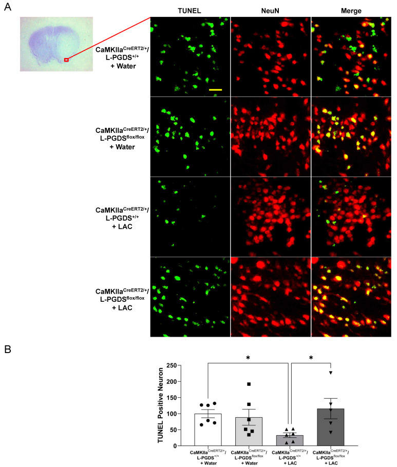
Ischemic stroke is one of the leading causes of permanent disability and death in adults worldwide. Apoptosis is a major element contributing to post-ischemic neuronal death. We previously found that low-dose alcohol consumption (LAC) protects against neuronal apoptosis in the peri-infarct cortex following transient focal cerebral ischemia. Lipocalin-type prostaglandin D2 synthase (L-PGDS), which is mainly localized in the central nervous system (CNS), was previously shown to inhibit neuronal apoptosis. Therefore, we determined whether L-PGDS is involved in the protective effect of LAC against post-ischemic neuronal apoptosis. Wild-type (WT), CaMKIIαCreERT2/+/L-PGDS+/+, and CaMKIIαCreERT2/+/L-PGDSflox/flox mice on a C57BL/6J background were gavage fed with ethanol or volume-matched water once a day for 8 weeks. Tamoxifen (2 mg/day) was given intraperitoneally to CaMKIIαCreERT2/+/L-PGDS+/+ and CaMKIIαCreERT2/+/L-PGDSflox/flox mice for 5 days during the fourth week. AT-56 (30 mg/kg/day), a selective inhibitor of L-PGDS, was given orally to AT-56-treated WT mice from the fifth week for four weeks. Cerebral ischemia/reperfusion (I/R) injury, TUNEL-positive neurons, and cleaved caspase-3-positive neurons were measured at 24 h of reperfusion after a 90 min unilateral middle cerebral artery occlusion (MCAO). We found that 0.7 g/kg/day but not 2.8 g/kg/day ethanol significantly upregulated L-PGDS in the cerebral cortex. In addition, 0.7 g/kg/day ethanol diminished cerebral ischemia/reperfusion (I/R) injury and TUNEL-positive and cleaved caspase-3-positive neurons in the peri-infarct cortex in WT and CaMKIIαCreERT2/+/L-PGDS+/+ mice. Furthermore, the neuroprotective effect of 0.7 g/kg/day ethanol was alleviated in AT-56-treated WT and CaMKIIαCreERT2/+/L-PGDSflox/flox mice. Our findings suggest that LAC may protect against cerebral I/R injury by suppressing post-ischemic neuronal apoptosis via an upregulated L-PGDS.

Keywords: L-PGDS; apoptosis; brain; ethanol; ischemic stroke.


Conflict of interest statement

The authors declare no conflict of interest.

Figures

Figure 1
Figure 1
Effect of chronic ethanol consumption on protein expression of L-PGDS in the cerebral cortex. (A) Representative Western blots. (B) Values are means ± SEM for 4–5 mice in each group. * p < 0.05. Analyzed using one-way ANOVA with Dunnett’s post hoc test.
Figure 2
Figure 2
Effect of AT-56 on cerebral I/R injury. (A) Representative brain sections stained with cresyl violet. (B) Total infarct volume. (C) Neurological deficit score. Values are means ± SEM for 4 mice in each group. * p < 0.05. ** p < 0.005. Analyzed using two-way ANOVA followed by Tukey’s test.
Figure 2
Figure 2
Effect of AT-56 on cerebral I/R injury. (A) Representative brain sections stained with cresyl violet. (B) Total infarct volume. (C) Neurological deficit score. Values are means ± SEM for 4 mice in each group. * p < 0.05. ** p < 0.005. Analyzed using two-way ANOVA followed by Tukey’s test.
Figure 3
Figure 3
Effect of forebrain neuron-specific L-PGDS knockdown on cerebral I/R injury. (A) PCR-based genotyping. The homozygous (fl/fl) mice with Cre positive were considered to be forebrain neuron-specific L-PGDS knockdown mice following tamoxifen treatment. (B) Western blots of L-PGDS in the cerebral cortex of tamoxifen-treated CaMKIIαCreERT2/+/L-PGDS+/+ and CaMKIIαCreERT2/+/L-PGDSflox/flox mice. (C) Representative brain sections stained with cresyl violet. (D) Total infarct volume. (E) Neurological deficit score. Values are means ± SEM for 5–6 mice in each group. * p < 0.05. ** p < 0.005. Analyzed using two-way ANOVA followed by Tukey’s test.
Figure 4
Figure 4
Effect of AT-56 on TUNEL-positive neurons in the cerebral cortex following transient focal cerebral ischemia. (A) Representative double staining of NeuN and TUNEL in the peri-infarct cortex of the temporal lobe at the section 0.23 mm caudal to bregma. Scale bar = 20 μm. (B) Values of TUNEL-positive neurons are means ± SEM for 4 mice in each group. * p < 0.05. Analyzed using two-way ANOVA followed by Tukey’s test.
Figure 5
Figure 5
Effect of forebrain neuron-specific L-PGDS knockdown on TUNEL-positive neurons in the cerebral cortex following transient focal cerebral ischemia. (A) Representative double staining of NeuN and TUNEL in the peri-infarct cortex of the temporal lobe at the section 0.23 mm caudal to bregma. Scale bar = 20 μm. (B) Values of TUNEL-positive neurons are means ± SEM for 5–6 mice in each group. * p < 0.05. Analyzed using two-way ANOVA followed by Tukey’s test.
Figure 6
Figure 6
Effect of AT-56 on cleaved caspase-3-positive neurons in the cerebral cortex following transient focal cerebral ischemia. (A) Representative double staining of NeuN and cleaved caspase-3 in the peri-infarct cortex of the parietal lobe at the section 0.23 mm caudal to bregma. Scale bar = 100 μm. (B) Values of cleaved caspase-3-positive neurons are means ± SEM for 4 mice in each group. * p < 0.05. Analyzed using two-way ANOVA followed by Tukey’s test.
Figure 7
Figure 7
Effect of forebrain neuron-specific L-PGDS knockdown on the cleaved caspase-3-positive neuron in the cerebral cortex following transient focal cerebral ischemia. (A) Representative double staining of NeuN and TUNEL in the peri-infarct cortex of the parietal lobe at the section 0.23 mm caudal to bregma. Scale bar = 100 μm. (B) Values of cleaved caspase-3-positive neurons are means ± SEM for 5–6 mice in each group. * p < 0.05. Analyzed using two-way ANOVA followed by Tukey’s test.

Similar articles

Cited by

References

    1. Benjamin E.J., Blaha M.J., Chiuve S.E., Cushman M., Das S.R., Deo R., de Ferranti S.D., Floyd J., Fornage M., Gillespie C., et al. Heart Disease and Stroke Statistics-2017 Update: A Report From the American Heart Association. Circulation. 2017;135:e146–e603. doi: 10.1161/CIR.0000000000000485. - DOI - PMC - PubMed
    1. Favate A.S., Younger D.S. Epidemiology of Ischemic Stroke. Neurol. Clin. 2016;34:967–980. doi: 10.1016/j.ncl.2016.06.013. - DOI - PubMed
    1. Jean W.C., Spellman S.R., Nussbaum E.S., Low W.C. Reperfusion injury after focal cerebral ischemia: The role of inflammation and the therapeutic horizon. Neurosurgery. 1998;43:1382–1396; discussion 1396–1397. doi: 10.1227/00006123-199812000-00076. - DOI - PubMed
    1. Kalogeris T., Baines C.P., Krenz M., Korthuis R.J. Cell biology of ischemia/reperfusion injury. Int. Rev. Cell Mol. Biol. 2012;298:229–317. - PMC - PubMed
    1. Elmore S. Apoptosis: A review of programmed cell death. Toxicol. Pathol. 2007;35:495–516. doi: 10.1080/01926230701320337. - DOI - PMC - PubMed

MeSH terms