Comparative Study
. 2021 Dec 28;23(1):299.
doi: 10.3390/ijms23010299.

Comparative Study of Two-Dimensional (2D) vs. Three-Dimensional (3D) Organotypic Kertatinocyte-Fibroblast Skin Models for Staphylococcus aureus (MRSA) Infection

Affiliations
Comparative Study

Comparative Study of Two-Dimensional (2D) vs. Three-Dimensional (3D) Organotypic Kertatinocyte-Fibroblast Skin Models for Staphylococcus aureus (MRSA) Infection

Nilakshi Barua et al. Int J Mol Sci. .

Abstract

The invasion of skin tissue by Staphylococcus aureus is mediated by mechanisms that involve sequential breaching of the different stratified layers of the epidermis. Induction of cell death in keratinocytes is a measure of virulence and plays a crucial role in the infection progression. We established a 3D-organotypic keratinocyte-fibroblast co-culture model to evaluate whether a 3D-skin model is more effective in elucidating the differences in the induction of cell death by Methicillin-resistant Staphylococcus aureus (MRSA) than in comparison to 2D-HaCaT monolayers. We investigated the difference in adhesion, internalization, and the apoptotic index in HaCaT monolayers and our 3D-skin model using six strains of MRSA representing different clonal types, namely, ST8, ST30, ST59, ST22, ST45 and ST239. All the six strains exhibited internalization in HaCaT cells. Due to cell detachment, the invasion study was limited up to two and a half hours. TUNEL assay showed no significant difference in the cell death induced by the six MRSA strains in the HaCaT cells. Our 3D-skin model provided a better insight into the interactions between the MRSA strains and the human skin during the infection establishment as we could study the infection of MRSA in our skin model up to 48 h. Immunohistochemical staining together with TUNEL assay in the 3D-skin model showed co-localization of the bacteria with the apoptotic cells demonstrating the induction of apoptosis by the bacteria and revealed the variation in bacterial transmigration among the MRSA strains. The strain representing ST59 showed maximum internalization in HaCaT cells and the maximum cell death as measured by Apoptotic index in the 3D-skin model. Our results show that 3D-skin model might be more likely to imitate the physiological response of skin to MRSA infection than 2D-HaCaT monolayer keratinocyte cultures and will enhance our understanding of the difference in pathogenesis among different MRSA strains.

Keywords: 3D skin model; HaCaT; MRSA; skin.


Conflict of interest statement

The authors declare no conflict of interest.

Figures

Figure 1
Figure 1
Adherence (A) and Internalization (B) of six MRSA strains representative of common ST types on HaCaT cells. Error bars represent SD of the mean, and statistical significance was calculated using (*** p-value < 0.001) one-way ANOVA with post hoc Tukey tests. The p-values are coded with respect to each strain and are derived from the comparison between the means of the two respective strains. $$$ (p < 0.001) shows the p-value of the respective strain when compared to ST8, ϕ (p < 0.05), and ϕϕϕ (p < 0.001) show significant difference when compared to ST30. ψ (p < 0.05) and ψψ (p < 0.01) depict the significant difference of the respective strain when compared to ST59. The columns with respect to the strains that do not have p-value symbols depict that there is no significant difference between the mean values with respect to each other. The MRSA ST8 was compared against each respective strain; ST30, ST22, ST45 and ST239 revealed p < 0.001 ($$$). For ST30 and ST59, p < 0.001 (ϕϕϕ), and for ST59 with ST239, p < 0.01 (ψψ), respectively. In (B), ST59 compared with ST45 and ST239 gave p < 0.05 (ψ), and for ST30 compared with ST239, p < 0.05 (ϕ).
Figure 2
Figure 2
A schematic diagram showing the development of the three-dimensional human organotypic keratinocyte-fibroblast co-culture model. The figure was produced using Servier Medical Art (http://smart.servier.com/ accessed on 1 November 2021) (A). Timeline of the development and infection of the organotypic keratinocyte-fibroblast co-culture model (B). H&E staining of the cultured organotypic keratinocyte-fibroblast co-culture model. The image represents Day 7 culture in air-liquid interface. Scale Bar shows 20 µm (C).
Figure 3
Figure 3
H&E stain of histological section of skin model at 48 h after inoculation with MRSA strain ST8 (A), ST30 (B), ST59 (C), ST22 (D), ST45 (E) and ST239 (F), respectively. White arrows show microcolonies of bacteria. Scale Bar depicts 50 µm.
Figure 4
Figure 4
Enumeration of adherent and internalized bacteria of MRSA strains representative of common ST types on skin model. Error bars represent SD of the mean, statistical significance was calculated using two-way ANOVA (*** p < 0.001) and Tukey post hoc test. The p-values are coded with respect to each strain and are derived from the comparison between the means of the two respective strains. p-values were significant at p < 0.05 when compared to ST30 (ϕ), ST59 (ψ), ST22 (θ), and ST45 (#).
Figure 5
Figure 5
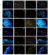
Images of a double-labelling of a cross-section of the skin model after 48 h infection with MRSA strain ST8 (A), ST30 (B), ST59 (C), ST22 (D), ST45 (E) and ST239 (F) using (i) DNA-binding Hoecsht stain (blue) to reveal the nuclei of the keratinocytes at the strata basale and spinosum, (ii) bacteria detected using anti-S. aureus antibody and Alexa Fluor® 568 conjugated secondary antibody, (iii) the Click-iT® TUNEL Alexa Fluor® 488 cell. (iv) is an overlay of the emission signals. It shows co-localization of bacteria and apoptosis/DNA damage in keratinocytes in strata spinosum. White hashed line demarcates the dermal epidermal boundary between the stratum basale and the collagen gel populated with fibroblasts. The yellow line in (i) shows the stratum spinosum and applies across (i) to (iv). The yellow arrows in (ii) show the bacteria in the collagen gel. The yellow circles in (iv) show the exfoliation of the skin model. Scale Bar shows 50 µm and applies to (i) to (iv).
Figure 6
Figure 6
Quantification of the apoptotic index (%) (A) in HaCaT cells 2 and half hour post infection and (B) in the skin model 48 h post infection with six MRSA strains representative of the common ST types. Error bars represent SD of the mean; statistical significance was calculated using one-way ANOVA (*** p < 0.001) and Tukey post hoc test. The p values are coded with respect to each strain and are derived from the comparison between the means of the two respective strains. Statistical significance was obtained at p < 0.05 when the strain was compared to ST8 ($), ST30 (ϕ) and ST59 (ψ), respectively.

Similar articles

Cited by

References

    1. DeLeo F.R., Otto M., Kreiswirth B.N., Chambers H.F. Community-associated meticillin-resistant Staphylococcus aureus. Lancet. 2010;375:1557–1568. doi: 10.1016/S0140-6736(09)61999-1. - DOI - PMC - PubMed
    1. Wong J.W.H., Ip M., Tang A., Wei V.W.I., Wong S.Y.S., Riley S., Read J.M., Kwok K.O. Prevalence and risk factors of community-associated methicillin-resistant staphylococcus aureus carriage in asia-pacific region from 2000 to 2016: A systematic review and meta-analysis. Clin. Epidemiol. 2018;10:1489–1501. doi: 10.2147/CLEP.S160595. - DOI - PMC - PubMed
    1. Sapugahawatte D.N., Li C., Yeoh Y.K., Dharmaratne P., Zhu C., Ip M. Swine methicillin-resistant Staphylococcus aureus carrying toxic-shock syndrome toxin gene in Hong Kong, China. Emerg. Microbes Infect. 2020;9:1534–1536. doi: 10.1080/22221751.2020.1785335. - DOI - PMC - PubMed
    1. Monecke S., Coombs G., Shore A.C., Coleman D.C., Akpaka P., Borg M., Chow H., Ip M., Jatzwauk L., Jonas D., et al. A field guide to pandemic, epidemic and sporadic clones of methicillin-resistant Staphylococcus aureus. PLoS ONE. 2011;6:e17936. doi: 10.1371/journal.pone.0017936. - DOI - PMC - PubMed
    1. Talan D.A., Krishnadasan A., Gorwitz R.J., Fosheim G.E., Limbago B., Albrecht V., Moran G.J. Comparison of staphylococcus aureus from skin and soft-tissue infections in us emergency Department patients, 2004 and 2008. Clin. Infect. Dis. 2011;53:144–149. doi: 10.1093/cid/cir308. - DOI - PubMed

Publication types