

Structural and Functional Change in Albino Rat Retina Induced by Various Visible Light Wavelengths
- PMID: 35008736
- PMCID: PMC8745104
- DOI: 10.3390/ijms23010309
Structural and Functional Change in Albino Rat Retina Induced by Various Visible Light Wavelengths
Abstract
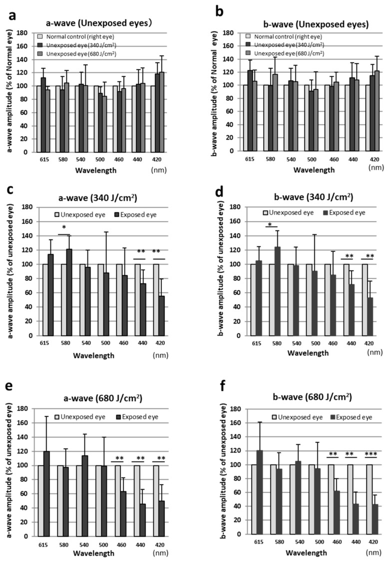
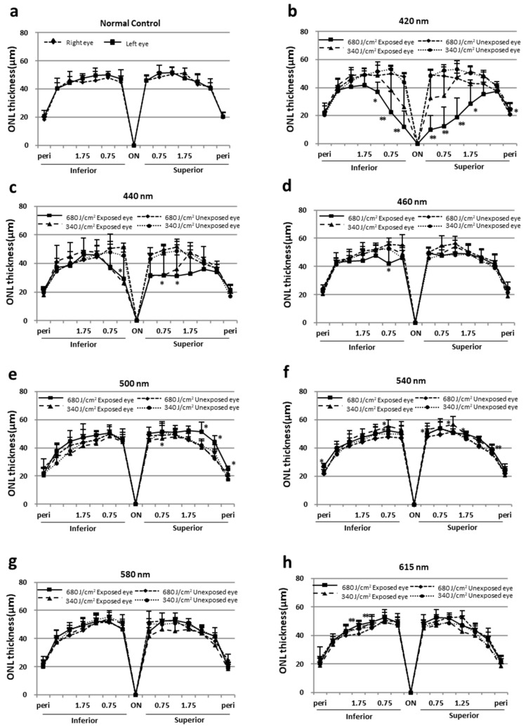
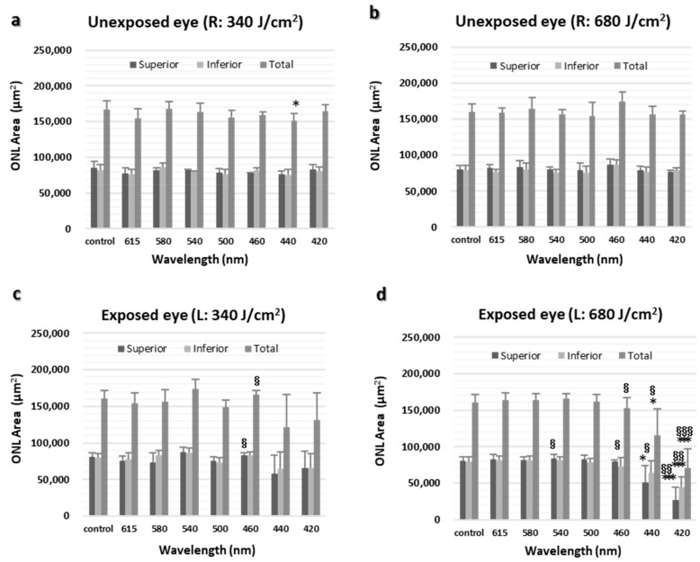
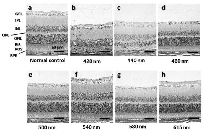
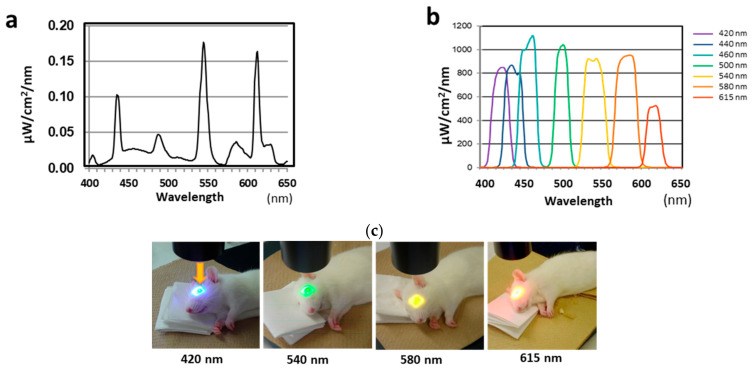
The effects of visible light, from short to long wavelengths, on the retina were investigated functionally and histologically. The left eyes of Sprague-Dawley albino rats (6-weeks old, n = 6 for each wavelength) were exposed to seven narrow-band wavelengths (central wavelengths, 421, 441, 459, 501, 541, 581, and 615 nm) with bandwidths of 16 to 29 nm (half bandwidth, ±8-14.5 nm) using a xenon lamp source with bandpass filters at the retinal radiant exposures of 340 and 680 J/cm2. The right unexposed eyes served as controls. Seven days after exposure, flash electroretinograms (ERGs) were recorded, and the outer nuclear layer (ONL) thickness was measured. Compared to the unexposed eyes, significant reductions in the a- and b-wave ERG amplitudes were seen in eyes exposed to 460-nm or shorter wavelengths of light. The ONL thickness near the optic nerve head also tended to decrease with exposure to shorter wavelengths. The decreased ERG amplitudes and ONL thicknesses were most prominent in eyes exposed to 420-nm light at both radiant exposures. When the wavelengths were the same, the higher the amount of radiant exposure and the stronger the damage. Compared to the unexposed eyes, the a- and b-waves did not decrease significantly in eyes exposed to 500-nm or longer wavelength light. The results indicate that the retinal damage induced by visible light observed in albino rats depends on the wavelength and energy level of the exposed light.
Keywords: electroretinograms; outer nuclear layer; retinal degeneration; visible light.
Conflict of interest statement
The authors declare no conflict of interest.
Figures
Similar articles
-
Protective effects of soft acrylic yellow filter against blue light-induced retinal damage in rats.Exp Eye Res. 2006 Dec;83(6):1493-504. doi: 10.1016/j.exer.2006.08.006. Epub 2006 Sep 25. Exp Eye Res. 2006. PMID: 16997296
-
Protective effect of TEMPOL derivatives against light-induced retinal damage in rats.Invest Ophthalmol Vis Sci. 2007 Apr;48(4):1900-5. doi: 10.1167/iovs.06-1057. Invest Ophthalmol Vis Sci. 2007. PMID: 17389526
-
Temporary elevation of the intraocular pressure by cauterization of vortex and episcleral veins in rats causes functional deficits in the retina and optic nerve.Exp Eye Res. 2003 Jul;77(1):27-33. doi: 10.1016/s0014-4835(03)00089-7. Exp Eye Res. 2003. PMID: 12823985
-
Functional evaluation of retina and optic nerve in the rat model of chronic ocular hypertension.Exp Eye Res. 2004 Jul;79(1):75-83. doi: 10.1016/j.exer.2004.02.011. Exp Eye Res. 2004. PMID: 15183102
-
Protective Effect of Highly Polymeric A-Type Proanthocyanidins from Seed Shells of Japanese Horse Chestnut (Aesculus turbinata BLUME) against Light-Induced Oxidative Damage in Rat Retina.Nutrients. 2018 May 10;10(5):593. doi: 10.3390/nu10050593. Nutrients. 2018. PMID: 29748512 Free PMC article.
Cited by
-
Mingmu Xiaoyao granules regulate the PI3K/Akt/mTOR signaling pathway to reduce anxiety and depression and reverse retinal abnormalities in rats.Front Pharmacol. 2022 Oct 5;13:1003614. doi: 10.3389/fphar.2022.1003614. eCollection 2022. Front Pharmacol. 2022. PMID: 36278192 Free PMC article.
References
-
- Noell W.K., Walker V.S., Kang B.S., Berman S. Retinal damage by light in rats. Investig. Ophthalmol. 1966;5:450–473. - PubMed
-
- Rapp L.M., Smith S.C. Morphologic comparisons between rhodopsin-mediated and short-wavelength classes of retinal light damage. Investig. Ophthalmol. Vis. Sci. 1992;33:3367–3377. - PubMed
MeSH terms
LinkOut - more resources
Full Text Sources
