. 2021 Dec 28;23(1):314.
doi: 10.3390/ijms23010314.

Availability of Arg, but Not tRNA, Is a Rate-Limiting Factor for Intracellular Arginylation

Affiliations

Availability of Arg, but Not tRNA, Is a Rate-Limiting Factor for Intracellular Arginylation

Irem Avcilar-Kucukgoze et al. Int J Mol Sci. .

Abstract

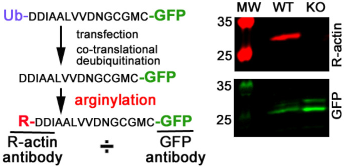
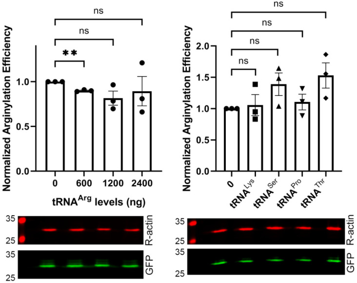
Protein arginylation, mediated by arginyltransferase ATE1, is a posttranslational modification of emerging biological importance that consists of transfer of the amino acid Arg from tRNA to protein and peptide targets. ATE1 can bind tRNA and exhibits specificity toward particular tRNA types, but its dependence on the availability of the major components of the arginylation reaction has never been explored. Here we investigated key intracellular factors that can potentially regulate arginylation in vivo, including several tRNA types that show strong binding to ATE1, as well as availability of free Arg, in an attempt to identify intracellular rate limiting steps for this enzyme. Our results demonstrate that, while modulation of tRNA levels in cells does not lead to any changes in intracellular arginylation efficiency, availability of free Arg is a potentially rate-limiting factor that facilitates arginylation if added to the cultured cells. Our results broadly outline global pathways that may be involved in the regulation of arginylation in vivo.

Keywords: arginine; arginine metabolism; arginylation; arginyltrasferase; tRNA.


Conflict of interest statement

The authors declare no conflict of interest.

Figures

Figure 1
Figure 1
tRNA and 18S rRNA inhibit arginylation in vitro. (A) Incorporation of 3H-Arg into angiotensin II in a typical arginylation reaction (typical), with and without addition of different RNA preparations, as marked on the figure. (B) Comparison of native tRNAArg expressed in E. coli and in vitro transcribed tRNAArg in the in vitro arginylation reaction. (C) Incorporation of 3H-Arg into angiotensin II in a typical arginylation reaction (typical), with and without addition of different tRNA types, as marked on the figure. Structural models of a typical tRNA (modeled from mouse tRNAThrACG-1-1) and the 18S rRNA fragment used in the reaction are shown on top for comparison of the predicted stem-loop structures. Cpm counts in each reaction set were normalized to the typical reaction. Error bars represent SEM, at least n = 3 independent reactions; *** p < 0.001, ns, not significant, Welch’s t-test.
Figure 2
Figure 2
Arginylation sensor for detection of intracellular arginylation. Left, diagram; right, representative Western blot of the arginylation sensor transfected into wild-type (WT) and Ate1 knockout (KO) mouse embryonic fibroblasts and visualized with antibodies to arginylated β actin (R-actin) and GFP. Ratio of the R-actin:GFP signal was used in the intracellular assays described below.
Figure 3
Figure 3
Transfection of cells with different tRNA species does not significantly affect intracellular arginylation. Charts (top) and representative Western blot images (bottom) of sensor-based quantification of arginylation in cells transfected with different amounts of tRNAArg (left) or 1200 ng of different tRNA species as indicated (right). Error bars represent SEM, n = 3 independent transfections; ** p < 0.01; ns, not significant; Welch’s t-test.
Figure 4
Figure 4
Addition of free Arg to the tissue culture medium increases the efficiency of arginylation. Chart (left) and representative Western blot image (right) of sensor-based quantification of arginylation in cells grown in Arg-free media supplemented with different amounts of Arg, as indicated. Error bars represent SEM; n = 4 independent transfections; * p < 0.05, ns, not significant, Welch’s t-test.
Figure 5
Figure 5
Addition of free Arg or tRNAArg to cells does not affect the levels of ATE1. Charts (top) and representative Western blot images (bottom) of sensor-based quantification of arginylation in cells grown in media supplemented with different amounts of Arg (left) or transfected with different amounts of tRNAArg (right). Error bars represent SEM, at least n = 3 independent transfections; ns, not significant, Welch’s t-test.
Figure 6
Figure 6
Arginine is a central component in a number of key physiological pathways. Major pathways that critically depend on Arg levels in the cell are shown. Alterations in Arg availability has the potential to critically affect the balance of these pathways, with important consequences to the cell.

Similar articles

References

    1. Wang J., Han X., Wong C.C., Cheng H., Aslanian A., Xu T., Leavis P., Roder H., Hedstrom L., Yates J.R., et al. Arginyltransferase ATE1 Catalyzes Midchain Arginylation of Proteins at Side Chain Carboxylates In Vivo. Chem. Biol. 2014;21:331–337. doi: 10.1016/j.chembiol.2013.12.017. - DOI - PMC - PubMed
    1. Wang J., Pejaver V.R., Dann G.P., Wolf M.Y., Kellis M., Huang Y., Garcia B.A., Radivojac P., Kashina A. Target site specificity and in vivo complexity of the mammalian arginylome. Sci. Rep. 2018;8:16177. doi: 10.1038/s41598-018-34639-6. - DOI - PMC - PubMed
    1. Cornachione A.S., Leite F.S., Wang J., Leu N.A., Kalganov A., Volgin D., Han X., Xu T., Cheng Y.-S., Yates J.R., et al. Arginylation of Myosin Heavy Chain Regulates Skeletal Muscle Strength. Cell Rep. 2014;8:470–476. doi: 10.1016/j.celrep.2014.06.019. - DOI - PMC - PubMed
    1. Saha S., Wong C.C., Xu T., Namgoong S., Zebroski H., Yates J.R., Kashina A. Arginylation and Methylation Double Up to Regulate Nuclear Proteins and Nuclear Architecture In Vivo. Chem. Biol. 2011;18:1369–1378. doi: 10.1016/j.chembiol.2011.08.019. - DOI - PMC - PubMed
    1. Rai R., Wong C.C.L., Xu T., Leu N.A., Dong D.W., Guo C., McLaughlin K.J., Yates J.R., III, Kashina A. Arginyltransferase regulates alpha cardiac actin function, myofibril formation and contractility during heart development. Development. 2008;135:3881–3889. doi: 10.1242/dev.022723. - DOI - PMC - PubMed

LinkOut - more resources