. 2021 Dec 28;23(1):318.
doi: 10.3390/ijms23010318.

Evaluation of the Relationships between Intestinal Regional Lymph Nodes and Immune Responses in Viral Infections in Children

Affiliations

Evaluation of the Relationships between Intestinal Regional Lymph Nodes and Immune Responses in Viral Infections in Children

Yayoi Aoki et al. Int J Mol Sci. .

Abstract

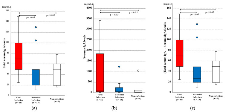
Viral infections increase the risk of developing allergies in childhood, and disruption of mucosal homeostasis is presumed to be involved. However, no study has reported a role for viral infections in such disruption. In this study, we clarified the mechanism of immunoglobulin A (IgA) overproduction in viral infections. Autopsies were performed on 33 pediatric cases, IgA and interferon (IFN)β levels were measured, and histopathological and immunohistochemical examinations were conducted. Furthermore, we cultured human cells and measured IFNβ and IgA levels to examine the effect of viral infections on IgA production. Blood IgA levels in viral infections were higher than in bacterial infections. Moreover, IFNβ levels in most viral cases were below the detection limit. Cell culture revealed increased IgA in gastrointestinal lymph nodes, especially in Peyer's patches, due to enhanced IFNβ after viral stimulation. Conversely, respiratory regional lymph nodes showed enhanced IgA with no marked change in IFNβ. Overproduction of IgA, identified as an aberration of the immune system and resulting from excessive viral infection-induced IFNβ was observed in the intestinal regional lymph nodes, particularly in Peyer's patches. Further, increased IgA without elevated IFNβ in the respiratory system suggested the possibility of a different mechanism from the gastrointestinal system.

Keywords: Peyer’s patch; cell culture; child; immune system; immunoglobulin A; interferon β; regional lymph node; viral infection.


Conflict of interest statement

The authors declare no conflict of interest.

Figures

Figure 1
Figure 1
(a) Total serum IgA levels, (b) serum sIgA levels, and (c) total serum IgA levels–serum sIgA levels in autopsy cases by infection category. The lines in the graphs indicate median values and the lines above and below the boxes show 90% confidence intervals. The arrows indicate significant differences between two groups, at p < 0.05 according to the Mann–Whitney U test.
Figure 2
Figure 2
Histopathology of pulmonary hilar lymph nodes and intestinal Peyer’s patches in (a) non-infection, (b) bacterial infection, and (c) viral infection autopsy cases. Pulmonary hilar lymph node: (i) HE staining (×100), (ii) IFNβ immunostaining (×100), (iii) IgA immunostaining (×100). Intestinal Peyer’s patches: (iv) HE staining (×100), (v) IFNβ immunostaining (×100), (vi) IgA immunostaining (×100).
Figure 2
Figure 2
Histopathology of pulmonary hilar lymph nodes and intestinal Peyer’s patches in (a) non-infection, (b) bacterial infection, and (c) viral infection autopsy cases. Pulmonary hilar lymph node: (i) HE staining (×100), (ii) IFNβ immunostaining (×100), (iii) IgA immunostaining (×100). Intestinal Peyer’s patches: (iv) HE staining (×100), (v) IFNβ immunostaining (×100), (vi) IgA immunostaining (×100).
Figure 3
Figure 3
(a) Changes in IFNβ levels after addition of poly(I:C) to intestinal lymph node-related cultured cells: (i) Peyer’s patch interstitial cells, (ii) intestinal tract membrane lymph node-origin interstitial cells, (iii) fossa axillaris lymph node-origin interstitial cells (control). (b) Changes in IFNβ levels after addition of poly(I:C) to respiratory lymph node-related cultured cells by concentration: (i) Pulmonary hilar node-origin interstitial cells, (ii) inguinal lymph node-origin interstitial cells (control). * Indicates a significant difference between the groups, at p < 0.05 by the Games–Howell test.
Figure 4
Figure 4
(a) Changes in IgA levels after addition of IFNβ to intestinal lymph node-related cultured cells: (i) Intestinal Peyer’s patch lymphocytes, (ii) mesenteric lymph node lymphocytes, (iii) fossa axillaris lymph node lymphocytes (control). (b) Changes in IgA levels after addition of IFNβ to respiratory lymph node-related cultured cells by concentration: (i) Pulmonary hilar lymph node lymphocytes, (ii) inguinal lymph node lymphocytes (control). * Indicates a significant difference between the groups, at p < 0.05 by the Games–Howell test.
Figure 5
Figure 5
Cultured intestinal Peyer’s patch lymphocytes (a) before and (b) after the addition of 1000 ng/mL IFNβ.
Figure 6
Figure 6
Cell sizes after the addition of IFNβ to lymphocytes in each type of regional lymph node. IFNβ concentration-dependent blast formation was observed in intestinal Peyer’s patch lymphocytes and pulmonary hilar lymph node lymphocytes.

Similar articles

Cited by

References

    1. Sevelsted A., Stokholm J., Bønnelykke K., Bisgaard H. Cesarean section and chronic immune disorders. Pediatrics. 2015;135:e92–e98. doi: 10.1542/peds.2014-0596. - DOI - PubMed
    1. Cheung D.S., Grayson M.H. Role of viruses in the development of atopic disease in pediatric patients. Curr. Allergy Asthma Rep. 2012;12:613–620. doi: 10.1007/s11882-012-0295-y. - DOI - PMC - PubMed
    1. Ebisawa M., Nishima S., Ohnishi H., Kondo N. Pediatric allergy and immunology in Japan. Pediatr. Allergy Immunol. 2013;24:704–714. doi: 10.1111/pai.12117. - DOI - PubMed
    1. Tantilipikorn P., Auewarakul P. Airway allergy and viral infection. Asian Pac. J. Allergy Immunol. 2011;29:113–119. - PubMed
    1. Castillo J.R., Peters S.P., Busse W.W. Asthma exacerbations: Pathogenesis, prevention, and treatment. J. Allergy Immunol. Pract. 2017;5:918–927. doi: 10.1016/j.jaip.2017.05.001. - DOI - PMC - PubMed

MeSH terms