. 2021 Dec 28;23(1):331.
doi: 10.3390/ijms23010331.

Extracellular Vesicles Mediate Communication between Endothelial and Vascular Smooth Muscle Cells

Affiliations

Extracellular Vesicles Mediate Communication between Endothelial and Vascular Smooth Muscle Cells

Marie Fontaine et al. Int J Mol Sci. .

Abstract

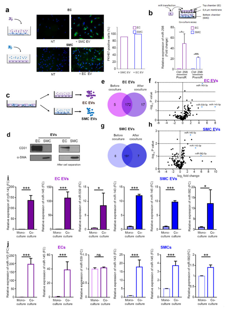
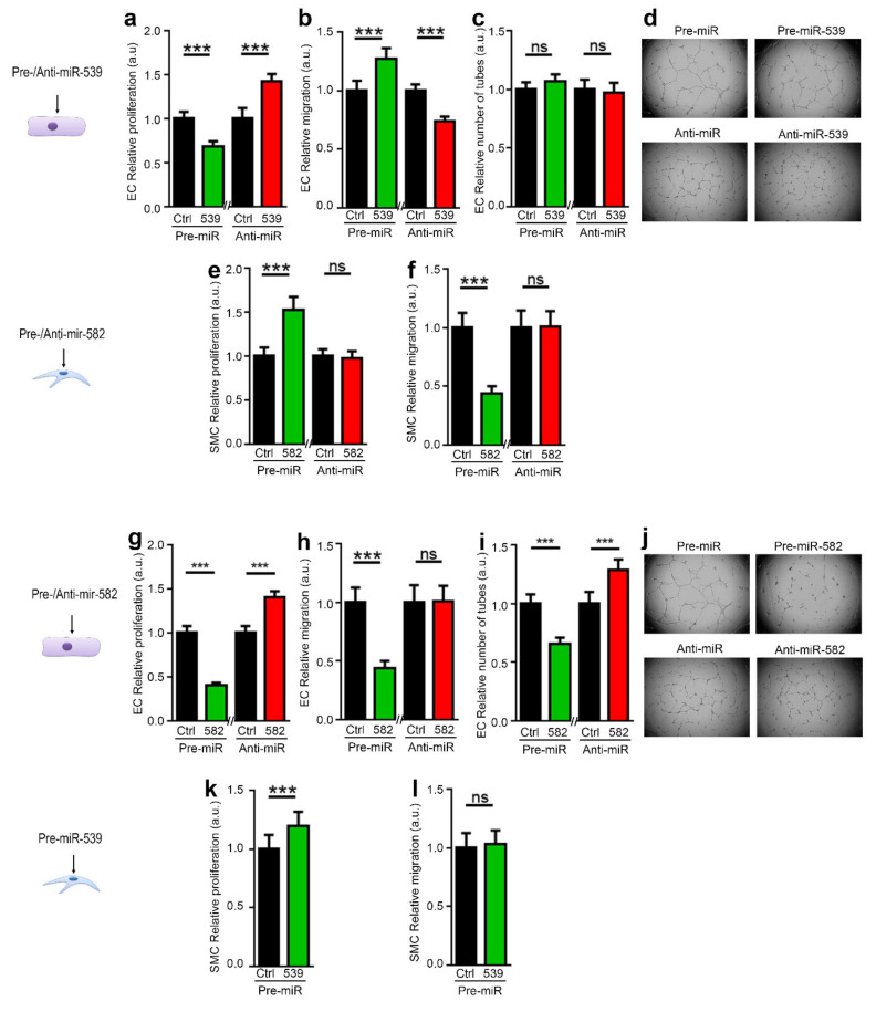
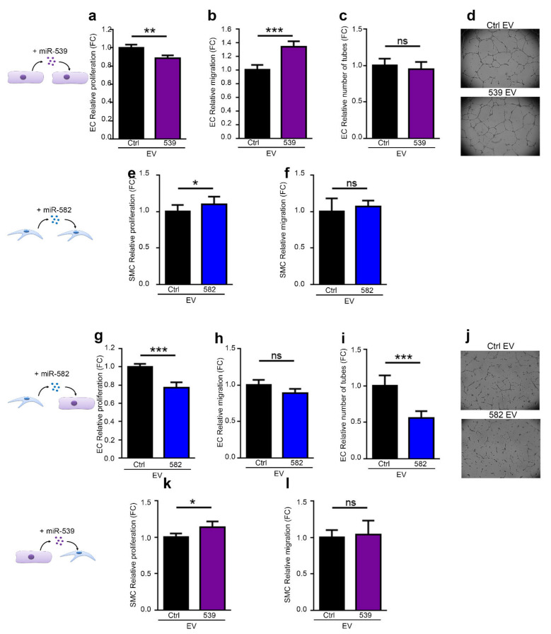
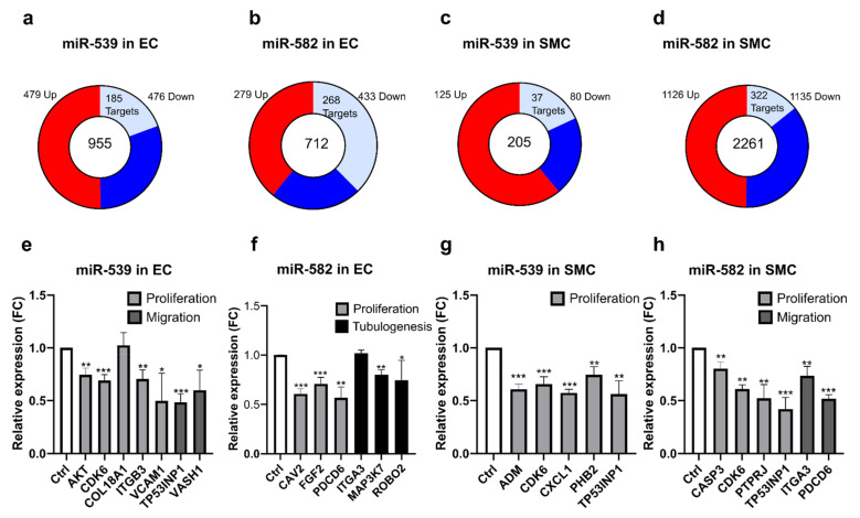
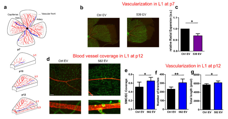
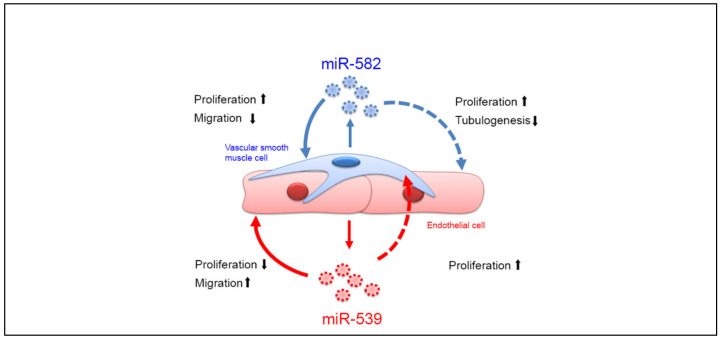
The recruitment of pericytes and vascular smooth muscle cells (SMCs) that enwrap endothelial cells (ECs) is a crucial process for vascular maturation and stabilization. Communication between these two cell types is crucial during vascular development and in maintaining vessel homeostasis. Extracellular vesicles (EVs) have emerged as a new communication tool involving the exchange of microRNAs between cells. In the present study, we searched for microRNAs that could be transferred via EVs from ECs to SMCs and vice versa. Thanks to a microRNA profiling experiment, we found that two microRNAs are more exported in each cell type in coculture experiments: while miR-539 is more secreted by ECs, miR-582 is more present in EVs from SMCs. Functional assays revealed that both microRNAs can modulate both cell-type phenotypes. We further identified miR-539 and miR-582 targets, in agreement with their respective cell functions. The results obtained in vivo in the neovascularization model suggest that miR-539 and miR-582 might cooperate to trigger the process of blood vessel coverage by smooth muscle cells in a mature plexus. Taken together, these results are the first to highlight the role of miR-539 and miR-582 in angiogenesis and communication between ECs and SMCs.

Keywords: angiogenesis; exosome; extracellular vesicle; miR-539; miR-582; microRNA.


Conflict of interest statement

The authors declare no conflict of interest.

Figures

Figure 1
Figure 1
ECs and SMCs communicate via EVs. (a) Fluorescence microscopy detection of the uptake of PHK67-labeled EVs (green) by ECs and SMCs (DAPI, blue), scale bars = 50 µm. Graph shows the percentage of positive cells for PKH67. (b) Overview of the experimental setup: miR-298 levels evaluated qPCR in SMCs cultured with ECs transfected with pre-miR-control or pre-miR-298 (blue bars). The purple bars show the reverse experiment in which the level of miR-298 was analyzed in EC cultured in presence of SMC transfected with pre-miR control or pre-miR-298. (c) Overview of the experimental setup. ECs and SMCs were cocultured for 48 h, separated using magnetic beads, and seeded as monoculture to produce EVs. microRNA content of cells and EVs were analyzed by microRNA qPCR profiling. (d) Western blot of CD31 and α-SMA in EVs isolated from EC and SMC cultures (left panels) or after coculture and separation of cells with CD31-coated magnetic bead (right panels) (e) Diagram of miRNAs common and specific to EC EVs before and after coculture. (f) Volcano plot of fold changes (log2 values) and probability values (−log10) for individual miRNAs in the EVs before and after coculture. (g) Diagram of miRNAs common and specific to SMC EVs before and after coculture. (h) Volcano plot of fold changes (log2 values) and probability values (−log10) for individual miRNAs in the SMC EVs before and after coculture. (i) Analyses of the expression of miR-143, miR-145, and miR-539 by qPCR in EC EVs before and after coculture with SMCs and the expression of miR-143, miR-145, and miR-582 in SMC EVs before and after coculture with ECs. (j) Analyses of the expression of miR-143, miR-145, and miR-539 by qPCR in ECs before and after coculture with SMCs and the expression of miR-143, miR-145, and miR-582 in SMCs before and after coculture with ECs. All data are the mean ± SEM (N = 3 independent experiments). * p < 0.05, ** p < 0.01, and *** p < 0.001 vs. the respective control.
Figure 2
Figure 2
miR-539 and miR-582 modulate EC and SMC functions in vitro. ECs were transfected with pre-miR-539 or a pre-miR-Control or with an anti-miR-control or an anti-miR-539 (ad). SMCs (e,f) or ECs (gj) were transfected with pre-miR-582 or a pre-miR-control or with an anti-miR-control or an anti-miR-582. (k,l) SMCs were transfected with pre-miR-539 or a pre-miR-control. Cells were then used in the following bioassays: (a,e,g,k) proliferation assay, (b,h) migration wound-healing assay of ECs, (l,f) migration of SMCs in boyden chamber assay, and (c,d,i,j) tubulogenesis assay of ECs. Right: representative images from d and j are shown. All data are the mean ± SD (n = 3 independent experiments). *** p < 0.001 vs. the respective control.
Figure 3
Figure 3
miR-539- and miR-582 can be transferred via EVs and modulate EC and SMC functions in vitro. EVs were purified from ECs transfected with pre-miR-539 (539 EV) or a pre-miR-Control (Ctrl EV) (a,b,k,l), or from SMCs transfected with pre-miR-582 (582 EV) on a pre-miR-control (Ctrl EVs) (ej), and used in the following bioassays, (a,g) EC proliferation, (b,h) migration wound-healing assay of ECs, and (c,d,i,j) tubulogenesis of ECs. Right: representative images from (c) and (i) are shown, (e,k) proliferation of SMCs, and (f,l) migration of SMCs in Boyden chamber assays. All data are the mean ± SD (n = 3 independent experiments). * p < 0.05, ** p < 0.01 and *** p < 0.001 vs. the respective control.
Figure 4
Figure 4
miR-539 and miR-582 regulate genes involved in angiogenesis-related biological processes in ECs and SMCs. (a,d) Transcriptomic analysis of ECs transfected with pre-miR-539 (a) and pre-miR-582 (b) and SMCs transfected with pre-miR-539 (c) and pre-miR-582 (d). Pie chart shows all significant regulated genes (p < 0.05) analyzed by RNA sequencing with the number of upregulated genes colored in red while downregulated genes are labeled in blue. Light blue shows number of potential microRNA targets. (eh) Validation of some putative targets by qPCR. (e) ECs were transfected with pre-miR-control or pre-miR-539. (f) ECs were transfected with pre-miR-control or pre-miR-582. (g) SMCs were transfected with pre-miR-control or pre-miR-539. (h) SMCs were transfected with pre-miR-control or pre-miR-582. All data are the mean ± SD (n = 3 independent experiments). * p < 0.05, ** p < 0.01, and *** p < 0.001 vs. the respective control.
Figure 5
Figure 5
miR-539 EVs and miR-582 EVs participate in vascular remodeling of the retina. (a) Cartoon illustrating the development of the superficial (L1), intermediate (L2), and deep vascular plexus (L3). (b) Left: retinal flat mount representation. For (bg), EVs were purified from ECs transfected with pre-miR-539 (539 EV) or a pre-miR-Control (Ctrl EV) or from SMCs transfected with pre-miR-582 (582 EV) on a pre-miR-control (Ctrl EV) (b) Isolectin-B4 staining on Postnatal Day 7 retinas from pups that were injected at Postnatal Day 1 with EVs. Scale bar: 25 µm. (fh). The histograms represent the quantification of the radial expansion from the optic nerve to the vascular front (c), N = 8 eyes, two independent experiments. (d) Confocal images of isolectin-B4/α -SMA staining on postnatal Day 12 retinas from pups that were injected at Postnatal Day 7 with miR-582 EVs in the superficial layer (L1). Scale bar: 200 µm. (eg). The histograms represent the quantification of the coverage of ECs by SMCs (Spearman’s rank correlation value) (e), the number of branches (f), and the total length of isolectin+ vessels (g) * p < 0.05, ** p = 0.01.
Figure 6
Figure 6
ECs and SMCs communicate via the exchange of microRNAs-loaded EVs. The coculture of ECs and SMCs induces a modification to the microRNAs’ cargo of EVs. After the coculture, ECs EVs are enriched in miR-539 while SMCs EVs are enriched in miR-582; then, cells exchange their EVs, inducing some modification to their cellular functions.

References

    1. Potente M., Gerhardt H., Carmeliet P. Basic and therapeutic aspects of angiogenesis. Cell. 2011;146:873–887. doi: 10.1016/j.cell.2011.08.039. - DOI - PubMed
    1. Lilly B. We have contact: Endothelial cell-smooth muscle cell interactions. Physiology. 2014;29:234–241. doi: 10.1152/physiol.00047.2013. - DOI - PubMed
    1. Gaengel K., Genové G., Armulik A., Betsholtz C. Endothelial-mural cell signaling in vascular development and angiogenesis. Arterioscler. Thromb. Vasc. Biol. 2009;29:630–638. doi: 10.1161/ATVBAHA.107.161521. - DOI - PubMed
    1. Sweeney M., Foldes G. It Takes Two: Endothelial-Perivascular Cell Cross-Talk in Vascular Development and Disease. Front. Cardiovasc. Med. 2018;5:154. doi: 10.3389/fcvm.2018.00154. - DOI - PMC - PubMed
    1. Mathieu M., Martin-Jaular L., Lavieu G., Théry C. Specificities of secretion and uptake of exosomes and other extracellular vesicles for cell-to-cell communication. Nat. Cell Biol. 2019;21:9–17. doi: 10.1038/s41556-018-0250-9. - DOI - PubMed

MeSH terms