. 2021 Dec 29;23(1):340.
doi: 10.3390/ijms23010340.

NLRP3 Inflammasome Activation Controls Vascular Smooth Muscle Cells Phenotypic Switch in Atherosclerosis

Affiliations

NLRP3 Inflammasome Activation Controls Vascular Smooth Muscle Cells Phenotypic Switch in Atherosclerosis

Fabienne Burger et al. Int J Mol Sci. .

Abstract

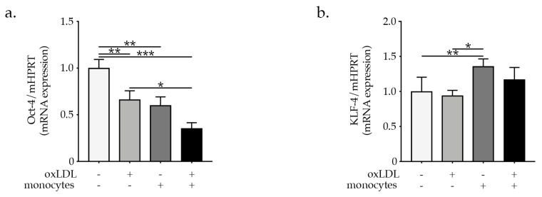
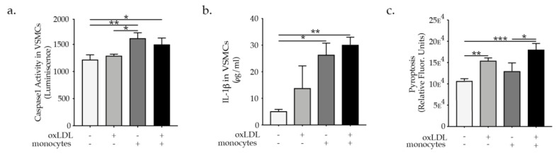
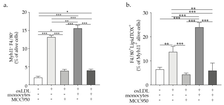
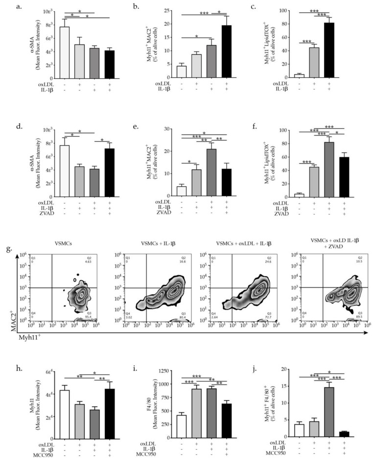
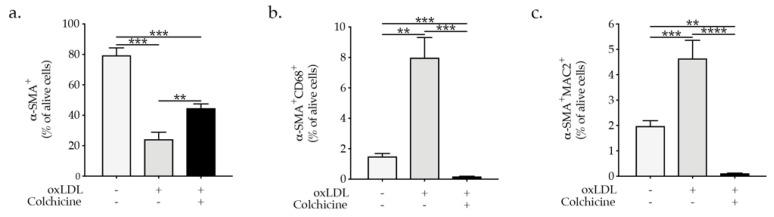
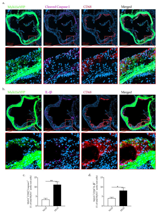
(1) Background: Monocytes and nucleotide-binding oligomerization domain-like receptor protein 3 (NLRP3) inflammasome orchestrate lipid-driven amplification of vascular inflammation promoting the disruption of the fibrous cap. The components of the NLRP3 inflammasome are expressed in macrophages and foam cells within human carotid atherosclerotic plaques and VSMCs in hypertension. Whether monocytes and NLRP3 inflammasome activation are direct triggers of VSMC phenotypic switch and plaque disruption need to be investigated. (2) Methods: The direct effect of oxLDL-activated monocytes in VSMCs co-cultured system was demonstrated via flow cytometry, qPCR, ELISA, caspase 1, and pyroptosis assay. Aortic roots of VSMCs lineage tracing mice fed normal or high cholesterol diet and human atherosclerotic plaques were used for immunofluorescence quantification of NLRP3 inflammasome activation/VSMCs phenotypic switch. (3) Results: OxLDL-activated monocytes reduced α-SMA, SM22α, Oct-4, and upregulation of KLF-4 and macrophage markers MAC2, F4/80 and CD68 expression as well as caspase 1 activation, IL-1β secretion, and pyroptosis in VSMCs. Increased caspase 1 and IL-1β in phenotypically modified VSMCs was detected in the aortic roots of VSMCs lineage tracing mice fed high cholesterol diet and in human atherosclerotic plaques from carotid artery disease patients who experienced a stroke. (4) Conclusions: Taken together, these results provide evidence that monocyte promote VSMC phenotypic switch through VSMC NLRP3 inflammasome activation with a likely detrimental role in atherosclerotic plaque stability in human atherosclerosis.

Keywords: NLRP3 inflammasome activation; atherosclerosis; atherosclerosis plaques stability; vascular smooth muscle; vascular smooth muscle phenotypic switch.


Conflict of interest statement

The authors declare no conflict of interest.

Figures

Figure 1
Figure 1
(a) Scheme of in vitro experimental setting and graph bars represent the mean ± SEM of flow cytometry analysis of VSMCs phenotypic switch (b) α-SMA+, (c) SM22α+ expressed as mean fluorescence intensity and (d) α-SMA+SM22α+, (e) MAC2+, (f) MAC2+F4/80+, and (g) CD68+ cells, expressed as percentage of indicated VSMC cells upon oxLDL treatment or co-culture with monocytes, with n = 6/group and * p  <  0.05, ** p  <  0.01, *** p  <  0.001, and **** p  <  0.0001, one-way ANOVA. Representative flow cytometry zebra plots of (h) α-SMA+SM22α+ and (i) MAC2+F4/80+ expression quantification.
Figure 2
Figure 2
Graph bars represent the mean ± SEM of mRNA expression of (a) Oct-4 and (b) KLF-4 indicating VSMC phenotypic switch upon oxLDL treatment or co-culture with monocytes or or oxLDL-activated monocytes, as indicated, with n = 6/group and * p < 0.05, ** p < 0.01, and *** p < 0.001, one-way ANOVA.
Figure 3
Figure 3
Graph’s bar represent the mean ± SEM of (a) caspase 1 activation, (b) IL-1β secretion, and (c) pyroptosis in VSMC upon oxLDL treatment or monocytes or oxLDL-activated monocytes supplementation as indicated, with n  =  6/group and * p  <  0.05, ** p  <  0.01, and *** p  <  0.001, one-way ANOVA.
Figure 4
Figure 4
Bars graphs represent the mean ± SEM of (a) Myh11+F4/80+-expressing VSMCs and (b) F4/80+LipidTOX+ (foam cells) formation as a percentage of alive cells as indicated upon oxLDL or monocytes, or oxLDL-activated monocytes supplementation in the presence or absence of MCC950, with n = 6/group and * p < 0.05, ** p < 0.01, *** p < 0.001, one-way ANOVA.
Figure 5
Figure 5
Graph bars represent the mean ± SEM of (a) α-SMA+, (b) Myh11+MAC2+ expression and (c) Myh11+LipidTOX+ (foam cells) formation of VSMCs upon oxLDL and IL-β treatment as indicated, with n  =  6/group and * p  < 0.05, ** p < 0.01, *** p  <  0.001. Graph bars represent the mean ± SEM of (d) α-SMA+, (e) Myh11+MAC2+, expressed as mean fluorescence intensity and (f) Myh11+LipidTOX+ (foam cells) expressed as a percentage of a alive VSMC upon oxLDL and/or IL-β and/or ZVAD treatment as indicated, with n  =  6/group and * p  <  0.05, ** p  <  0.01, *** p  <  0.001, one-way ANOVA. (g) Representative flow cytometry zebra plots of Myh11+MAC2+ cells. Graph bars represent the mean ± SEM of (h) Myh11+, (i) F4/80+ expressed as mean fluorescence intensity and (j) Myh11+F4/80+ as a percentage of alive VSMC upon oxLDL and/or IL-β and/or MCC950 treatment as indicated, with n  =  6/group and * p  <  0.05, ** p  <  0.01, *** p  <  0.001, one-way ANOVA.
Figure 6
Figure 6
Graph bars represent the mean ± SEM of (a) α-SMA+, (b) α-SMA+CD68+, and (c) α-SMA+MAC2+ expression in VSMCs upon colchicine and/or oxLDL treatment as indicated, with n  =  5–6/group and ** p  <  0.01, *** p  <  0.001, **** p  <  0.0001 one-way ANOVA.
Figure 7
Figure 7
Representative immunofluorescence staining of Myh11eYFP+ cells, expressing (a) cleaved caspase 1, CD68 and (b) IL-1β, CD68 in the aortic roots of Apoe−/− Myh11ERT2-CreR26R-eYFP mice showing NLRP3 inflammasome activation in VSMC undergoing phenotypic switch in vivo, taken by confocal microscopy (LSM 800 Airyscan). Graph bars show the mean ± SEM of (c) Myh11eYFP+ CD68+ Cleaved Caspase1+ and (d). Myh11eYFP+ CD68+ IL-1β co-expressing cells as a percentage of all Myh11eYFP+ cells in the aortic roots plaquesm with n = 8/group and * p  <  0.05, ** p  <  0.01, unpaired t-test.
Figure 8
Figure 8
Representative immunofluorescence staining of Myh11+ cells, expressing (a) cleaved caspase 1, CD68 and (b) IL-1β, CD68 in human atherosclerotic plaques associated with NLRP3-inflammasome activation in VSMC and linked to plaque rupture in human carotid artery disease shown by confocal microscopy. Graph bars show the mean ± SEM of (c) Myh11+ CD68+ cleaved caspase 1 + and (d) Myh11+ CD68+ IL-1β+ co-expressing cells as a percentage of plaque Myh11 cells+ cells with n  =  12/group and * p  <  0.05, unpaired t-test.

Similar articles

Cited by

References

    1. Şahin B., Ilgün G. Risk factors of deaths related to cardiovascular diseases in World Health Organization (WHO) member countries. Health Soc. Care Community. 2020;30:73–80. doi: 10.1111/hsc.13156. - DOI - PubMed
    1. Hansson G.K., Libby P., Tabas I. Inflammation and plaque vulnerability. J. Intern. Med. 2015;278:483–493. doi: 10.1111/joim.12406. - DOI - PMC - PubMed
    1. Gupta R.M., Lee-Kim V.S., Libby P. The March of Monocytes in Atherosclerosis. Circ. Res. 2020;126:1324–1326. doi: 10.1161/CIRCRESAHA.120.316981. - DOI - PMC - PubMed
    1. Woollard K.J., Geissmann F. Monocytes in atherosclerosis: Subsets and functions. Nat. Rev. Cardiol. 2010;7:77–86. doi: 10.1038/nrcardio.2009.228. - DOI - PMC - PubMed
    1. Jacinto T.A., Meireles G.S., Dias A.T., Aires R., Porto M.L., Gava A.L., Vasquez E.C., Pereira T.M.C., Campagnaro B.P., Meyrelles S.S. Increased ROS production and DNA damage in monocytes are biomarkers of aging and atherosclerosis. Biol. Res. 2018;51:33. doi: 10.1186/s40659-018-0182-7. - DOI - PMC - PubMed

MeSH terms