

DUSP-1 Induced by PGE2 and PGE1 Attenuates IL-1β-Activated MAPK Signaling, Leading to Suppression of NGF Expression in Human Intervertebral Disc Cells
- PMID: 35008797
- PMCID: PMC8745672
- DOI: 10.3390/ijms23010371
DUSP-1 Induced by PGE2 and PGE1 Attenuates IL-1β-Activated MAPK Signaling, Leading to Suppression of NGF Expression in Human Intervertebral Disc Cells
Abstract
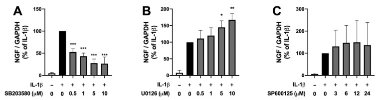
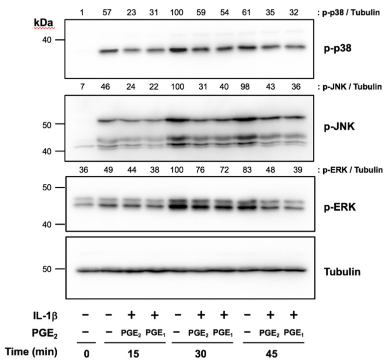
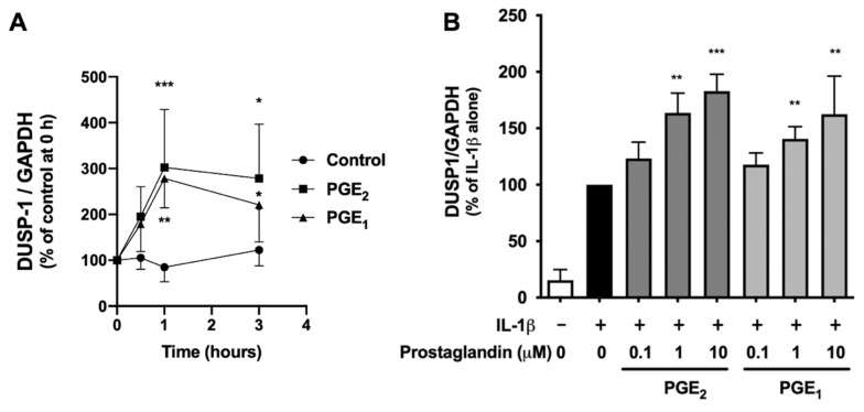
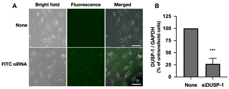
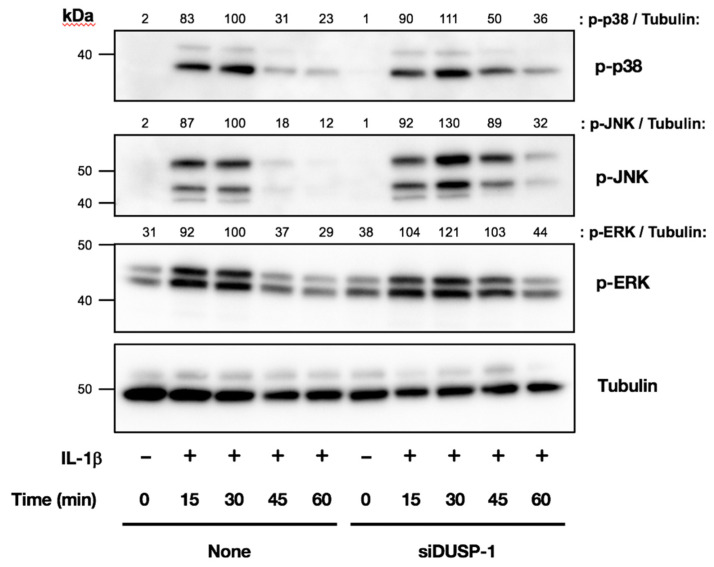
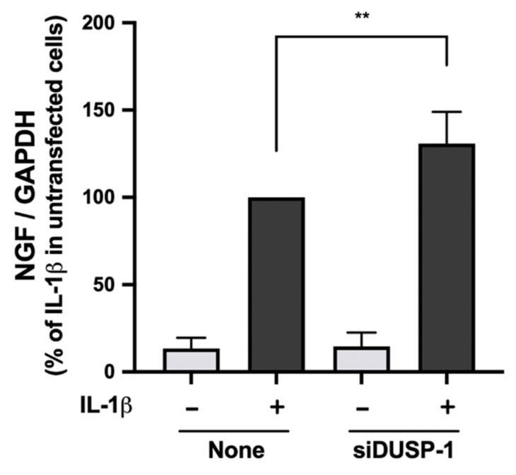
The molecular mechanism of discogenic low back pain (LBP) involves nonphysiological nerve invasion into a degenerated intervertebral disc (IVD), induced by nerve growth factor (NGF). Selective cyclooxygenase (COX)-2 inhibitors are mainly used in the treatment of LBP, and act by suppressing the inflammatory mediator prostaglandin E2 (PGE2), which is induced by inflammatory stimuli, such as interleukin-1β (IL-1β). However, in our previous in vitro study using cultured human IVD cells, we demonstrated that the induction of NGF by IL-1β is augmented by a selective COX-2 inhibitor, and that PGE2 and PGE1 suppress NGF expression. Therefore, in this study, to elucidate the mechanism of NGF suppression by PGE2 and PGE1, we focused on mitogen-activated protein kinases (MAPKs) and its phosphatase, dual-specificity phosphatase (DUSP)-1. IL-1β-induced NGF expression was altered in human IVD cells by MAPK pathway inhibitors. PGE2 and PGE1 enhanced IL-1β-induced DUSP-1 expression, and suppressed the phosphorylation of MAPKs in human IVD cells. In DUSP-1 knockdown cells established using small interfering RNA, IL-1β-induced phosphorylation of MAPKs was enhanced and prolonged, and NGF expression was significantly enhanced. These results suggest that PGE2 and PGE1 suppress IL-1β-induced NGF expression by suppression of the MAPK signaling pathway, accompanied by increased DUSP-1 expression.
Keywords: dual-specificity phosphatase (DUSP)-1; interleukin-1β (IL-1β); intervertebral disc (IVD); mitogen-activated protein kinase (MAPK); nerve growth factor (NGF); prostaglandin E1 (PGE1); prostaglandin E2 (PGE2).
Conflict of interest statement
The authors declare that they have no conflicts of interest associated with this study.
Figures
Similar articles
-
PGE1 Attenuates IL-1β-induced NGF Expression in Human Intervertebral Disc Cells.Spine (Phila Pa 1976). 2016 Jun;41(12):E710-E716. doi: 10.1097/BRS.0000000000001379. Spine (Phila Pa 1976). 2016. PMID: 26656048
-
Prostaglandin E2 induces dual-specificity phosphatase-1, thereby attenuating inflammatory genes expression in human osteoarthritic synovial fibroblasts.Prostaglandins Other Lipid Mediat. 2021 Jun;154:106550. doi: 10.1016/j.prostaglandins.2021.106550. Epub 2021 Apr 18. Prostaglandins Other Lipid Mediat. 2021. PMID: 33857603
-
Regulation of nerve growth factor by anti-inflammatory drugs, a steroid, and a selective cyclooxygenase 2 inhibitor in human intervertebral disc cells stimulated with interleukin-1.Spine (Phila Pa 1976). 2013 Aug 1;38(17):1466-72. doi: 10.1097/BRS.0b013e318294edb1. Spine (Phila Pa 1976). 2013. PMID: 23574818
-
Nerve growth factor promotes expression of novel genes in intervertebral disc cells that regulate tissue degradation: Laboratory investigation.J Neurosurg Spine. 2014 Oct;21(4):653-61. doi: 10.3171/2014.6.SPINE13756. Epub 2014 Jul 25. J Neurosurg Spine. 2014. PMID: 25062286
-
Nerve Growth Factor Is Regulated by Toll-Like Receptor 2 in Human Intervertebral Discs.J Biol Chem. 2016 Feb 12;291(7):3541-51. doi: 10.1074/jbc.M115.675900. Epub 2015 Dec 14. J Biol Chem. 2016. PMID: 26668319 Free PMC article.
Cited by
-
Osteoarthritis Pain.Int J Mol Sci. 2022 Apr 22;23(9):4642. doi: 10.3390/ijms23094642. Int J Mol Sci. 2022. PMID: 35563035 Free PMC article. Review.
-
Genomic Interplay between Neoneurogenesis and Neoangiogenesis in Carcinogenesis: Therapeutic Interventions.Cancers (Basel). 2023 Mar 16;15(6):1805. doi: 10.3390/cancers15061805. Cancers (Basel). 2023. PMID: 36980690 Free PMC article. Review.
-
Acetylation-regulated DUSP1 deficiency contributes to renal fibrosis progression.Theranostics. 2025 Mar 3;15(9):3781-3796. doi: 10.7150/thno.108992. eCollection 2025. Theranostics. 2025. PMID: 40213676 Free PMC article.
-
Concepts of Regeneration for Spinal Diseases in 2022.Int J Mol Sci. 2022 Aug 26;23(17):9710. doi: 10.3390/ijms23179710. Int J Mol Sci. 2022. PMID: 36077105 Free PMC article.
-
Network Pharmacology and In Vivo Experimental Verification of the Mechanism of the Qing'e Pill for Treating Intervertebral Disc Degeneration.Curr Comput Aided Drug Des. 2025;21(4):534-548. doi: 10.2174/0115734099356426241119051916. Curr Comput Aided Drug Des. 2025. PMID: 39623712 Free PMC article.
References
-
- Vos T., Abajobir A.A., Abbafati C., Abbas K.M., Abate K.H., Abd-Allah H., Abdulle A.M., Abebo T.A., Abera S.F., Aboyans V., et al. Global, regional, and national incidence, prevalence, and years lived with disability for 328 diseases and injuries for 195 countries, 1990–2016: A systematic analysis for the Global Burden of Disease Study 2016. Lancet. 2017;390:1211–1259. doi: 10.1016/S0140-6736(17)32154-2. - DOI - PMC - PubMed
MeSH terms
Substances
Grants and funding
LinkOut - more resources
Full Text Sources
Research Materials
Miscellaneous
