. 2021 Dec 30;23(1):394.
doi: 10.3390/ijms23010394.

Transport and Toxicity of Methylmercury-Cysteine in Cultured BeWo Cells

Affiliations

Transport and Toxicity of Methylmercury-Cysteine in Cultured BeWo Cells

Srividya Ganapathy et al. Int J Mol Sci. .

Abstract

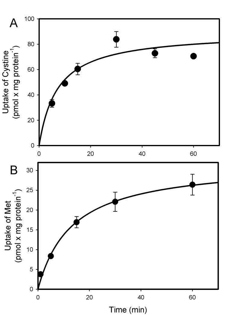
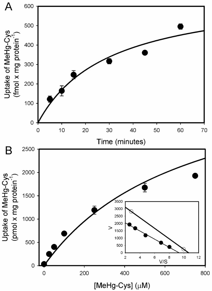
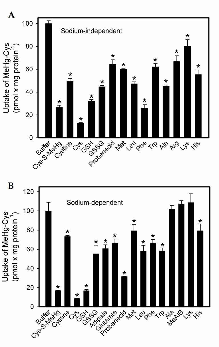
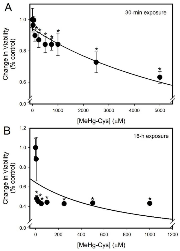
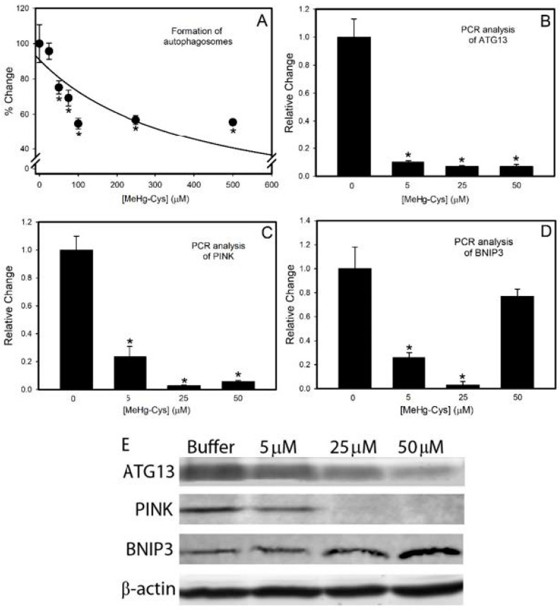
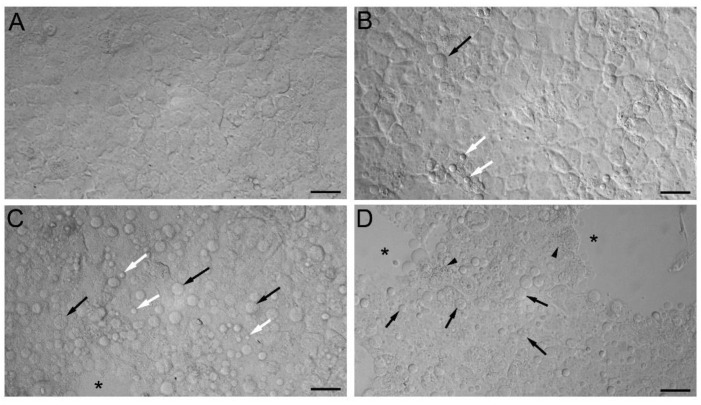
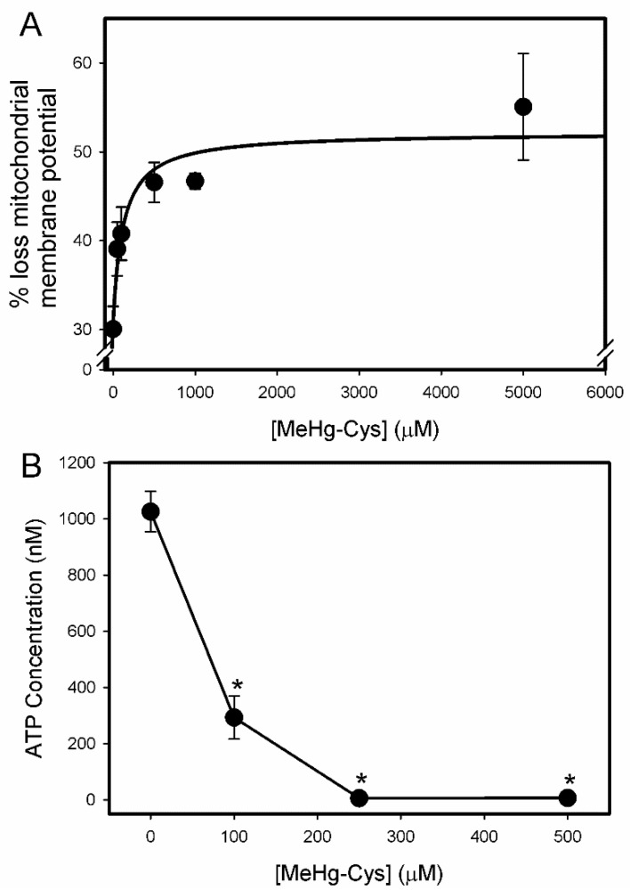
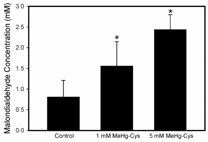
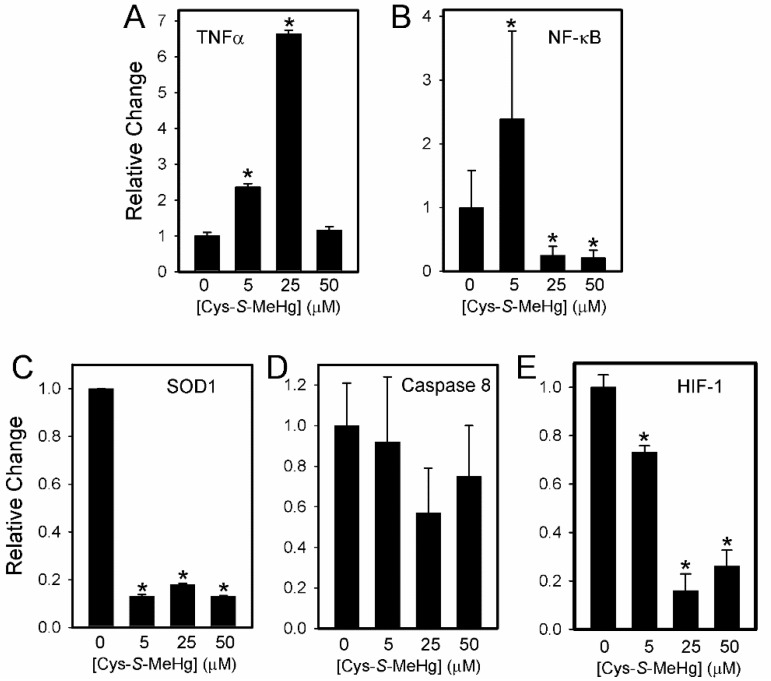
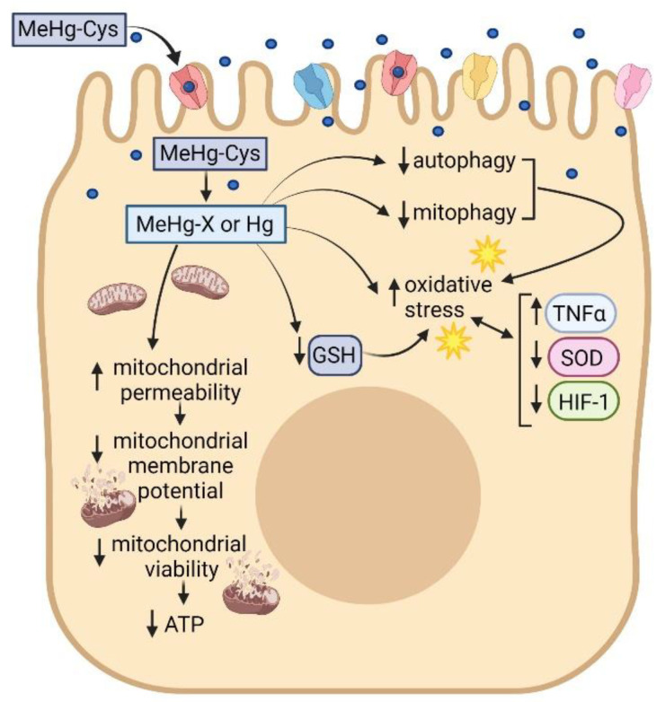
Mercury is a heavy metal toxicant that is prevalent throughout the environment. Organic forms of mercury, such as methylmercury (MeHg), can cross the placenta and can lead to lasting detrimental effects in the fetus. The toxicological effects of MeHg on the placenta itself have not been clearly defined. Therefore, the purpose of the current study was to assess the transport of MeHg into placental syncytiotrophoblasts and to characterize the mechanisms by which MeHg exerts its toxic effects. Cultured placental syncytiotrophoblasts (BeWo) were used for these studies. The transport of radioactive MeHg was measured to identify potential mechanisms involved in the uptake of this compound. The toxicological effects of MeHg on BeWo cells were determined by assessing visible pathological change, autophagy, mitochondrial viability, and oxidative stress. The findings of this study suggest that MeHg compounds are transported into BeWo cells primarily by sodium-independent amino acid carriers and organic anion transporters. The MeHg altered mitochondrial function and viability, decreased mitophagy and autophagy, and increased oxidative stress. Exposure to higher concentrations of MeHg inhibited the ability of cells to protect against MeHg-induced injury. The findings show that MeHg is directly toxic to syncytiotrophoblasts and may lead to disruptions in the fetal/maternal transfer of nutrients and wastes.

Keywords: autophagy; mercury; oxidative stress; placenta; syncytiotrophoblast; toxicology.


Conflict of interest statement

The authors declare no conflict of interest.

Figures

Figure 1
Figure 1
Uptake of 5 µM [35S]-cystine (A) and 5 µM [3H]-methionine (B) in BeWo cells. Uptake was carried out at 37 °C for various times between 5 and 60 min. Results are presented as mean ± SE. Data represent 3 experiments performed in triplicate.
Figure 2
Figure 2
Time course of 5 µM [14C]-MeHg-Cys uptake in BeWo cells (A). Uptake was carried out at 37 °C for time periods ranging from 5 to 60 min. Saturation kinetics for the transport of [14C]-MeHg-Cys (B) were assessed in BeWo cells (Inset: Eadie–Hofstee plot). Cells were incubated for 30 min with 5 µM [14C]-MeHg-Cys in the presence of unlabeled MeHg-Cys. Results are presented as mean ± SE. Data represent 3 experiments performed in triplicate.
Figure 3
Figure 3
Substrate specificity analyses of [14C]-MeHg-Cys transport in BeWo cells. Cells were incubated for 30 min at 37 °C with [14C]-MeHg-Cys in the presence of various unlabeled compounds (1 mM Cys-MeHg and cystine; 3 mM all others) under sodium-independent (A) and sodium-dependent (B) conditions. Results are presented as mean ± SE. Data represent 3 experiments performed in triplicate. *, significantly different (p < 0.05) from cells exposed to buffer only.
Figure 4
Figure 4
Cellular viability of BeWo cells exposed to MeHg-Cys. BeWo cells were exposed to various concentrations of MeHg-Cys for 30 min (A) or 16 h (B) at 37 °C and cell viability was measured using a methylthiazoletetrazolium (MTT) assay. The concentration of MeHg-Cys was reduced for the 16 h exposure because higher concentrations resulted in overt cell death. Results are presented as mean ± SE. Data represent 3 experiments performed in triplicate. *, significantly different (p < 0.05) from unexposed cells.
Figure 5
Figure 5
Indicators of autophagy in BeWo cells. BeWo cells were exposed to various concentrations of MeHg-Cys for 30 min at 37 °C, and the formation of autophagosomes was measured (A). For analyses of mRNA expression and protein levels, cells were exposed to MeHg-Cys for 16 h at 37 °C. RNA and protein were isolated from the control and exposed cells, and quantitative PCR (qPCR) was used to analyze levels of ATG13 (B), PINK (C), and BNIP3 (D). Western blotting (E) measured protein levels of ATG13 (72 kDa), PINK (63 kDa), and BNIP3 (22 kDa). β-actin (42 kDa) was used as the control. Results are presented as mean ± SE. Data represent 3 experiments performed in triplicate. *, significantly different (p < 0.05) from unexposed cells.
Figure 6
Figure 6
Morphological analyses of BeWo cells were exposed to MeHg-Cys. BeWo cells were exposed to buffer (A) or 100 µM (B), 250 µM (C), or 500 µM (D) MeHg-Cys for 30 min at 37 °C. Cells exposed to buffer (A) exhibited normal morphology while cells exposed to MeHg exhibited blebs (white arrows) and detaching cells (black arrows). Gaps (*) in the monolayer were evident in cells exposed to 250 and 500 µM MeHg-Cys. Small organelles (arrowheads) were identified in the cytoplasm of cells exposed to 500 µM MeHg-Cys. Images shown are representative of 3 experiments performed in triplicate. Scale bar = 40 µm.
Figure 7
Figure 7
Mitochondrial disruptions in BeWo cells exposed to MeHg-Cys. BeWo cells were exposed to buffer or various concentrations of MeHg-Cys for 30 min at 37 °C. The loss of mitochondrial membrane potential (A) was measured using fluorescence-activated cell sorting (FACS). Changes in ATP levels were measured under the same exposure conditions (B). Results are presented as mean ± SE. Data represent 3 experiments performed in triplicate. *, significantly different (p < 0.05) from unexposed cells.
Figure 8
Figure 8
Lipid peroxidation in BeWo cells exposed to MeHg-Cys. BeWo cells were exposed to 1 mM or 5 mM MeHg-Cys for 30 min at 37 °C. Results are presented as mean ± SE. Data represent 3 experiments performed in triplicate. *, significantly different (p < 0.05) from unexposed cells.
Figure 9
Figure 9
Indicators of oxidative stress in BeWo cells exposed to MeHg-Cys. BeWo cells were exposed to buffer or 5 µM, 25 mM, or 50 µM MeHg-Cys for 16 h at 37 °C. RNA was isolated from the control and exposed cells and quantitative PCR was used to analyze the expression of TNFα (A), NF-κB (B), SOD1 (C), Caspase 8 (D), and HIF-1 (E). Results are presented as mean ± SE. Data represent 3 experiments performed in triplicate. *, significantly different (p < 0.05) from unexposed cells.
Figure 10
Figure 10
Uptake of MeHg-Cys by placental syncytiotrophoblasts leads to intracellular intoxication characterized by oxidative stress and mitochondrial dysfunction. This figure was created by BioRender.

References

    1. ATSDR, Agency for Toxic Substances and Disease Registry, Toxicological Profile for Mercury . U.S. Department of Health and Human Services. Public Health Service, Centers for Disease Control; Atlanta, GA, USA: 2008. [(accessed on 21 July 2021)]. Available online: https://www.atsdr.cdc.gov/toxprofiles/tp46.pdf.
    1. Donohue A., Wagner C.L., Burch J.B., Rothenberg S.E. Blood total mercury and methylmercury among pregnant mothers in Charleston, South Carolina, USA. J. Expo. Sci. Environ. Epidemiol. 2018;28:494–504. doi: 10.1038/s41370-018-0033-1. - DOI - PMC - PubMed
    1. Wickliffe J.K., Lichtveld M.Y., Zijlmans C.W., MacDonald-Ottevanger S., Shafer M., Dahman C., Harville E.W., Drury S., Landburg G., Ouboter P. Exposure to total and methylmercury among pregnant women in Suriname: Sources and public health implications. J. Expo. Sci. Environ. Epidemiol. 2020;31:117–125. doi: 10.1038/s41370-020-0233-3. - DOI - PMC - PubMed
    1. Llorente Ballesteros M.T., Garcia Barrado B., Navarro Serrano I., Izquierdo Alvarez S., Del Pueyo Garcia Anaya M., Gonzalez Munoz M.J. Evaluation of blood mercury and serum selenium levels in the pregnant population of the Community of Madrid, Spain. J. Trace Elements Med. Biol. 2020;57:60–67. doi: 10.1016/j.jtemb.2019.09.008. - DOI - PubMed
    1. Bridges C.C., Zalups R.K. Mechanisms involved in the transport of mercuric ions in target tissues. Arch. Toxicol. 2017;91:63–81. doi: 10.1007/s00204-016-1803-y. - DOI - PMC - PubMed

MeSH terms

LinkOut - more resources