. 2021 Dec 30;23(1):403.
doi: 10.3390/ijms23010403.

Clinical Significance of Stem Cell Biomarkers EpCAM, LGR5 and LGR4 mRNA Levels in Lymph Nodes of Colon Cancer Patients

Affiliations

Clinical Significance of Stem Cell Biomarkers EpCAM, LGR5 and LGR4 mRNA Levels in Lymph Nodes of Colon Cancer Patients

Manar AbdelMageed et al. Int J Mol Sci. .

Abstract

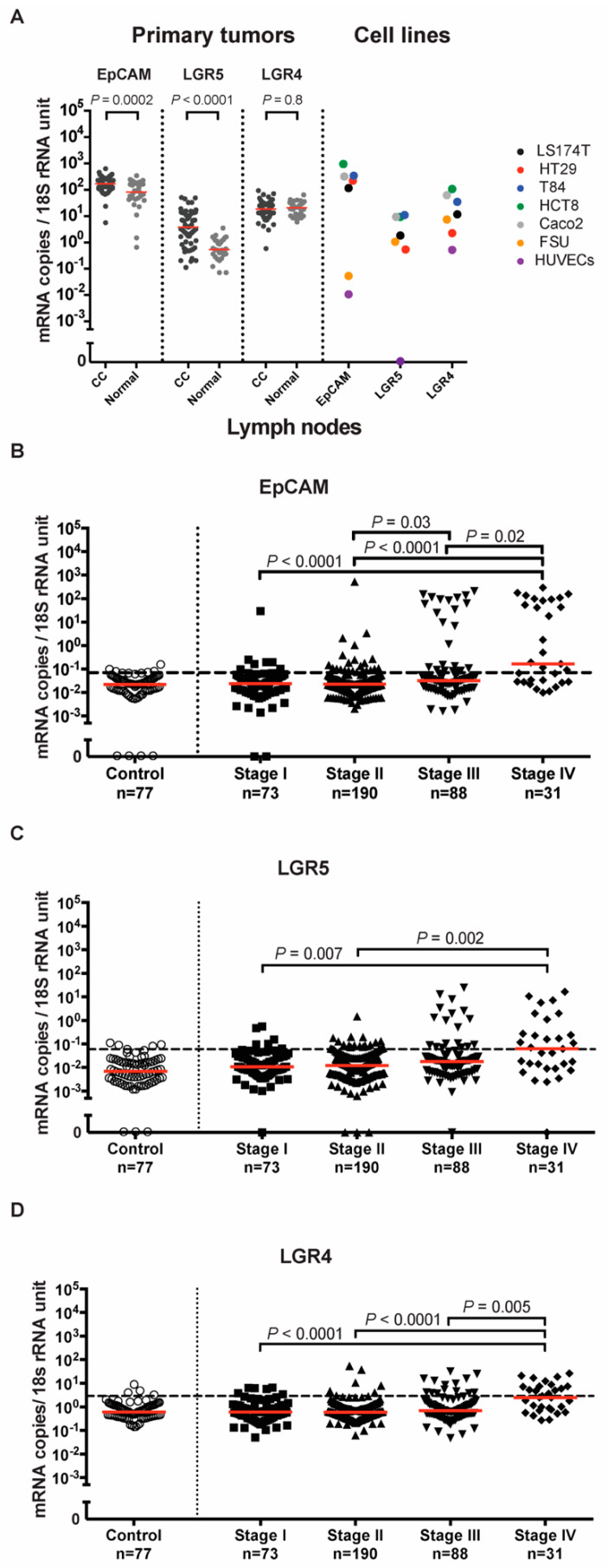
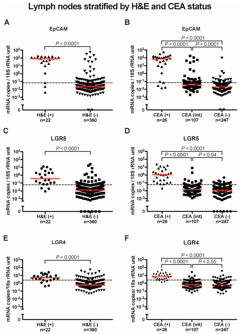
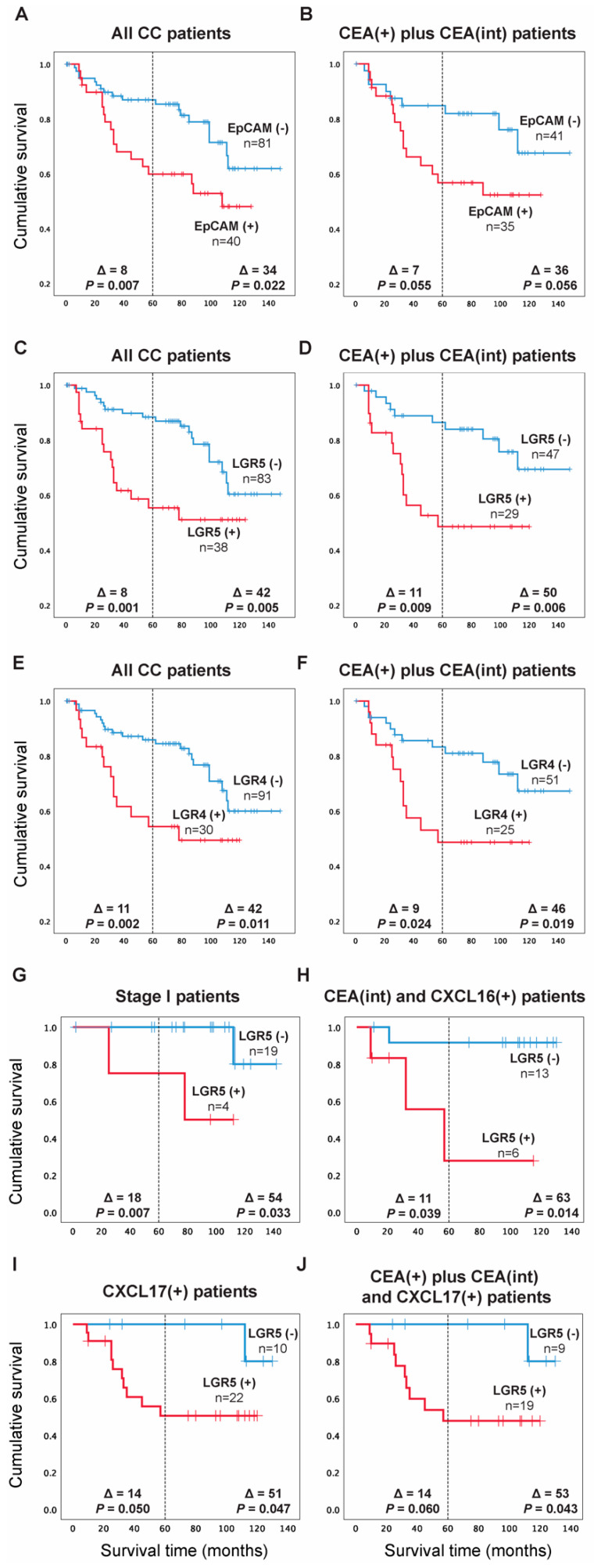
The significance of cancer stem cells (CSCs) in initiation and progression of colon cancer (CC) has been established. In this study, we investigated the utility of measuring mRNA expression levels of CSC markers EpCAM, LGR5 and LGR4 for predicting survival outcome in surgically treated CC patients. Expression levels were determined in 5 CC cell lines, 66 primary CC tumors and 382 regional lymph nodes of 121 CC patients. Prognostic relevance was determined using Kaplan-Meier survival and Cox regression analyses. CC patients with lymph nodes expressing high levels of EpCAM, LGR5 or LGR4 (higher than a clinical cutoff of 0.07, 0.06 and 2.558 mRNA copies/18S rRNA unit, respectively) had a decreased mean survival time of 32 months for EpCAM and 42 months for both LGR5 and LGR4 at a 12-year follow-up (p = 0.022, p = 0.005 and p = 0.011, respectively). Additional patients at risk for recurrence were detected when LGR5 was combined with the biomarkers CXCL17 or CEA plus CXCL16. In conclusion, the study underscores LGR5 as a particularly useful prognostic biomarker and illustrates the strength of combining biomarkers detecting different subpopulations of cancer cells and/or cells in the tumor microenvironment for predicting recurrence.

Keywords: CEA; CXCL16; CXCL17; EpCAM; LGR4; LGR5; colon cancer; prognosis; qRT-PCR; regional lymph nodes; stem cell markers.


Conflict of interest statement

The authors declare no competing interests.

Figures

Figure 1
Figure 1
EpCAM, LGR5, and LGR4 mRNA expression levels in tissues and cell lines. (A) mRNA expression levels in primary colon cancer (CC) tissues, resected normal colon tissues, and in a panel of colon cancer cell lines; LS174T, HT29, T84, HCT8 and Caco2, primary foreskin fibroblast cells (FSU) and endothelial cells (HUVECs). Red horizontal lines indicate median values. (BD) mRNA expression levels in lymph nodes from noncancerous disease patients (Control) and colon cancer patients in different TNM stages (Stage I–IV). Red horizontal lines indicate median values. Dashed horizontal lines indicate clinical cutoff values of 0.07, 0.06 and 2.558 mRNA copies/18S rRNA unit for EpCAM, LGR5 and LGR4, respectively. n = number of lymph nodes. p-values were calculated by two-tailed Mann-Whitney test in (A) and Kruskal-Wallis nonparametric ANOVA followed by post hoc Dunn’s test for multiple comparisons in (BD).
Figure 2
Figure 2
EpCAM, LGR5 and LGR4 mRNA expression in lymph nodes stratified by H&E and CEA status. (A) EpCAM, (C) LGR5, and (E) LGR4 mRNA levels in nonmetastatic (H&E(-)) and metastatic (H&E(+)) lymph nodes. In (B,D,F), lymph nodes were divided into three groups according to their CEA mRNA levels; CEA(-) = CEA mRNA levels < 0.013 copies/18S rRNA unit, CEA(int) = intermediate CEA mRNA levels, that is 0.013-3.67 copies/18S rRNA unit, and CEA(+) = CEA mRNA levels > 3.67 copies/18S rRNA unit. Red horizontal lines indicate median values. Dashed horizontal lines indicate clinical cutoff values of 0.07, 0.06 and 2.558 mRNA copies/18S rRNA unit for EpCAM, LGR5 and LGR4 respectively. n = number of lymph nodes. p-values were calculated by two-tailed Mann-Whitney test for comparison between expression levels in (A,C,E) and by Kruskal-Wallis nonparametric ANOVA followed by post hoc Dunn’s test for multiple comparisons in (B,D,F).
Figure 3
Figure 3
Two-color immunofluorescence staining of primary colon cancer tissue with anti-LGR5 and BerEP4, and anti-CEA and BerEP4. (A) Anti-LGR5, (E) Anti-CEA both red color. (B,F) BerEP4 mAb, green color. (C,G) Overlays giving yellow color of double-stained areas. (D) FITC-conjugated mouse IgG; negative control for BerEP4. (H) Rabbit IgG; negative control for anti-LGR5 and anti-CEA. Original magnification: ×200.
Figure 4
Figure 4
Kaplan-Meier cumulative survival curves for CC patients divided into two groups in (A) EpCAM(-) and EpCAM(+) according to the median of the expression level in the highest lymph nodes of the CC patients in TNM stages III and IV (0.07 mRNA copies/18S rRNA unit). (C) LGR5(-) and LGR5(+) according to the median of the expression level in lymph nodes of all CC patients in TNM stage IV (0.06 mRNA copies/18S rRNA unit). (E) LGR4(-) and LGR4(+) according to the 75th percentile of LGR4 mRNA expression values in all CC patients’ highest lymph nodes (2.558 mRNA copies/18S rRNA unit). In (B,D,F), the Kaplan-Meier cumulative survival curves for EpCAM, LGR5 and LGR4 patients are restricted to the CEA(+) plus CEA(int) subgroup of CC patients. In (GJ), the Kaplan-Meier cumulative survival curves for LGR5 patients are restricted to TNM stage I patients only (G), CEA(int) plus CXCL16(+) subgroups of patients (H), CXCL17(+) patients only (I) and CEA(+) plus CEA(int) plus CXCL17(+) patients subgroups (J). The patients were followed for 12 years. Differences in disease-free survival time after surgery between the two groups are given as a ∆-value in months and statistical significance as p-values. n = number of patients in the respective group.

References

    1. Yadav A.K., Desai N.S. Cancer stem cells: Acquisition, characteristics, therapeutic implications, targeting strategies and future prospects. Stem. Cell Rev. Rep. 2019;15:331–355. doi: 10.1007/s12015-019-09887-2. - DOI - PubMed
    1. Munro M.J., Wickremesekera S.K., Peng L., Tan S.T., Itinteang T. Cancer stem cells in colorectal cancer: A review. J. Clin. Pathol. 2018;71:110–116. doi: 10.1136/jclinpath-2017-204739. - DOI - PubMed
    1. Hatano Y., Fukuda S., Hisamatsu K., Hirata A., Hara A., Tomita H. Multifaceted interpretation of colon cancer stem cells. Int. J. Mol. Sci. 2017;18:1446. doi: 10.3390/ijms18071446. - DOI - PMC - PubMed
    1. Huang L., Yang Y., Yang F., Liu S., Zhu Z., Lei Z., Guo J. Functions of EpCAM in physiological processes and diseases (Review) Int. J. Mol. Med. 2018;42:1771–1785. doi: 10.3892/ijmm.2018.3764. - DOI - PMC - PubMed
    1. Van der Gun B.T.F., Melchers L.J., Ruiters M.H.J., de Leij L.F.M.H., McLaughlin P.M.J., Rots M.G. EpCAM in carcinogenesis: The good, the bad or the ugly. Carcinogenesis. 2010;31:1913–1921. doi: 10.1093/carcin/bgq187. - DOI - PubMed

MeSH terms

LinkOut - more resources