. 2021 Dec 31;23(1):431.
doi: 10.3390/ijms23010431.

Butein and Frondoside-A Combination Exhibits Additive Anti-Cancer Effects on Tumor Cell Viability, Colony Growth, and Invasion and Synergism on Endothelial Cell Migration

Affiliations

Butein and Frondoside-A Combination Exhibits Additive Anti-Cancer Effects on Tumor Cell Viability, Colony Growth, and Invasion and Synergism on Endothelial Cell Migration

Shahrazad Sulaiman et al. Int J Mol Sci. .

Abstract

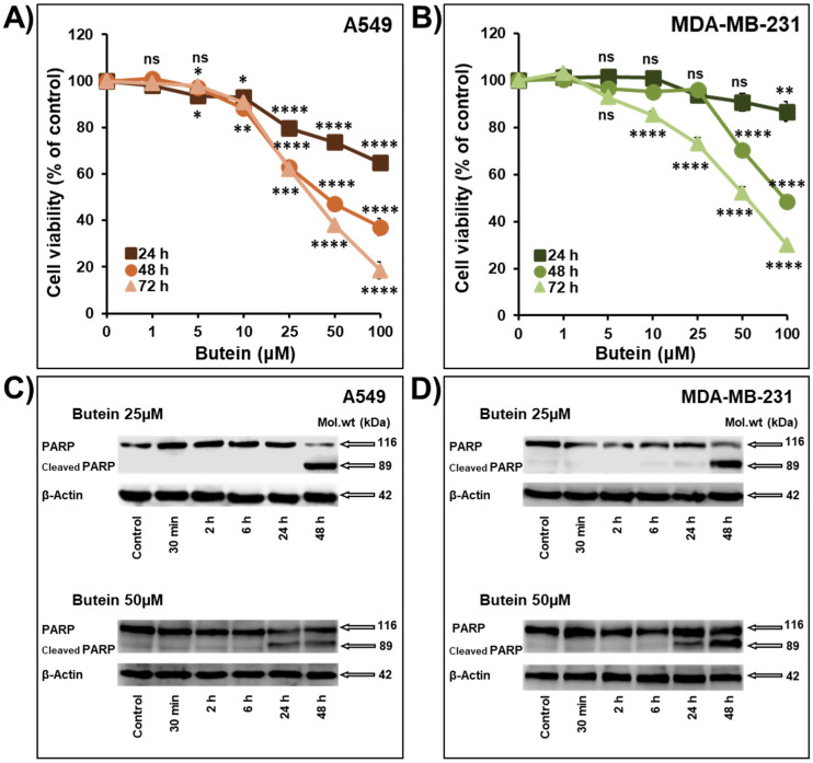
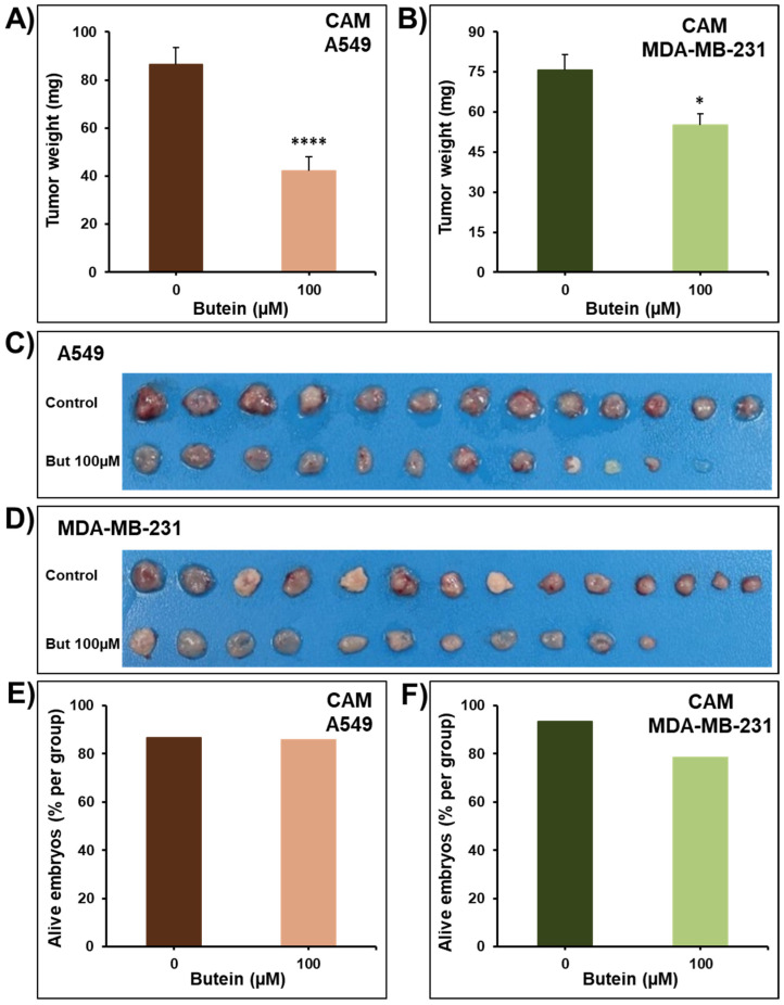
Despite the significant advances in targeted- and immuno-therapies, lung and breast cancer are at the top list of cancer incidence and mortality worldwide as of 2020. Combination therapy consisting of a mixture of different drugs taken at once is currently the main approach in cancer management. Natural compounds are extensively investigated for their promising anti-cancer potential. This study explored the anti-cancer potential of butein, a biologically active flavonoid, on two major solid tumors, namely, A549 lung and MDA-MB-231 breast cancer cells alone and in combination with another natural anti-cancer compound, frondoside-A. We demonstrated that butein decreases A549 and MDA-MB-231 cancer cell viability and colony growth in vitro in addition to tumor growth on chick embryo chorioallantoic membrane (CAM) in vivo without inducing any noticeable toxicity. Additionally, non-toxic concentrations of butein significantly reduced the migration and invasion of both cell lines, suggesting its potential anti-metastatic effect. We showed that butein anti-cancer effects are due, at least in part, to a potent inhibition of STAT3 phosphorylation, leading to PARP cleavage and consequently cell death. Moreover, we demonstrated that combining butein with frondoside-A leads to additive effects on inhibiting A549 and MDA-MB-231 cellular viability, induction of caspase 3/7 activity, inhibition of colony growth, and inhibition of cellular migration and invasion. This combination reached a synergistic effect on the inhibition of HUVECs migration in vitro. Collectively, this study provides sufficient rationale to further carry out animal studies to confirm the relevance of these compounds' combination in cancer therapy.

Keywords: STAT3; angiogenesis; breast cancer; butein; frondoside-A; invasion; lung cancer; tumor growth; viability.


Conflict of interest statement

The authors declare no conflict of interest. The funders had no role in the design of the study; in the collection, analyses, or interpretation of data; in the writing of the manuscript, or in the decision to publish the results.

Figures

Figure 1
Figure 1
Inhibition of cellular viability associated PARP cleavage by butein. Exponentially growing A549 (A) and MDA-MB-231 (B) cells were treated with vehicle (0.1% DMSO) and the indicated concentrations of butein for 24, 48, and 72 h. Viable cells were determined using the CellTiter-Glo Luminescent Cell Viability Assay, based on ATP quantification, which indicates the presence of viable cells. Experiments were repeated at least three times. Western blot analysis shows PARP cleavage after butein (25 and 50 μM) treatment in A549 (C) and MDA-MB-231 (D) cancer cells. β-actin was used as a loading control. The data shown are representative of three in-dependent experiments. Shapes represent means; bars represent S.E.M. * Significantly different at p < 0.05, ** Significantly different at p < 0.01, *** Significantly different at p < 0.001, **** Significantly different at p < 0.0001. ns—non-significant.
Figure 2
Figure 2
Effect of butein on colony growth. The growth of cancer cell-derived colonies from A549 (A) and MDA-MB-231 (B) cells was assessed by measuring the number of the colonies in control and butein-treated wells for seven days. (C,D) Representative pictures of the control and butein-treated colonies are shown for A549 and MDA-MB-231 cancer cells. Experiments were repeated at least three times. Columns represent means; bars represent S.E.M. * Significantly different at p < 0.05, ** Significantly different at p < 0.01, *** Significantly different at p < 0.001, **** Significantly different at p < 0.0001. ns—non-significant.
Figure 3
Figure 3
Impact of butein on tumor growth using the in vivo CAM tumor xenograft model. Volumes of 1 × 106 of A549 (A,C) and MDA-MB-231 (B,D) cells were grafted on the CAM of 9 days (E9) chick embryos. Tumors were treated with butein (100 µM) every 48 h for a total of 6 days. At E17, tumors were collected, weighed, and photographed (C,D). The viability of the chick embryos was assessed, and the percentage of alive embryos was determined (E,F). Columns are means; bars are S.E.M. * Significantly different at p < 0.05. **** Significantly different at p < 0.0001.
Figure 4
Figure 4
Butein impairs cancer cell migration and invasion. Wounds were introduced in A549 (A) and MDA-MB-231 (B) cells’ confluent monolayers cultured in the presence or absence (control) of butein (5 and 10 µM). The mean distance that cells travelled from the edge of the scraped area after 2 and 6 h was measured using an inverted microscope. A549 (C) and MDA-MB-231 (D) cells were incubated for 24 h in the presence or absence of butein (5 and 10 µM). Cells that invaded the Matrigel and crossed the 8 µm pores insert were determined using the CellTiter-Glo Luminescent Cell Viability Assay. All experiments were repeated at least three times. Columns or shapes represent means; bars represent S.E.M. * Significantly different at p < 0.05, ** Significantly different at p < 0.01, *** Significantly different at p < 0.001. ns—non-significant.
Figure 5
Figure 5
Western blot showing the inhibition of STAT3 phosphorylation by butein in A549 lung cancer cells (A,B), and MDA-MB-231 breast cancer cells (C,D). Effect of butein on total STAT3 (EH). Each cell line was treated with 25 and 50 μM butein, and proteins were extracted at the indicated time-points (0.5, 2, 6, 24, and 48 h). β-actin was used as a loading control. The data shown are representative of three independent experiments. Columns represent means; bars represent S.E.M. * Significantly different at p < 0.05, ** Significantly different at p < 0.01, *** Significantly different at p < 0.001, **** Significantly different at p < 0.0001. ns—non-significant.
Figure 6
Figure 6
Effect of butein in combination with frondoside-A on the inhibition of cell viability of A549 (A) and MDA-MB-231 cells (B) after 48 h treatment. Effect of combinations of butein and frondoside-A on cell viability compared with the calculated additive effects of the two drugs alone (C,D). Induction of caspase 3/7 activity was also analyzed in A549 and MDA-MB-231 cells treated for 48 h with frondoside-A (2.5 and 1 µM, respectively), butein (50 µM) and their combination (E,F). Effect of combinations of butein and frondoside-A on caspase 3/7 activity compared with the calculated additive effects of the two drugs alone (G,H). Data were normalized to the number of viable cells per well and expressed as fold induction compared to the control group. All experiments were repeated at least three times. Columns are means; bars are S.E.M. The statistical significance is compared to the control except for the specified lines. * Significantly different at p < 0.05, ** Significantly different at p < 0.01, *** Significantly different at p < 0.001, **** Significantly different at p < 0.0001. ns—non-significant.
Figure 7
Figure 7
Impact of butein in combination with frondoside-A on A549 colony growth after 14 days of treatment (A,B). Experiments were repeated three times. Columns represent means; bars represent S.E.M. The statistical significance is compared to the control except for the specified lines. ** Significantly different at p < 0.01, *** Significantly different at p < 0.001, **** Significantly different at p < 0.0001.
Figure 8
Figure 8
Effect of butein (25 µM) in combination with frondoside-A (0.5 µM) on HUVECs migration after 8 h of treatment (A). Effect of the combination on HUVECs migration compared with the calculated additive effects of the two drugs alone (B). Data were expressed as fold induction compared to the control group. (CE) Impact of butein and frondoside-A alone and in combination on HUVECs capillary-like-structure formation and cell viability after 8 h of treatment. The statistical significance is compared to the control (4% FBS) except for the specified lines. All experiments were repeated at least three times. Columns are means; bars are S.E.M. ** Significantly different at p < 0.01, *** Significantly different at p < 0.001, **** Significantly different at p < 0.0001. ns—non-significant.
Figure 9
Figure 9
Impact of butein combination with frondoside-A on cellular invasion. A549 (A) and MDA-MB-231 (B) cells were treated for 24 h with butein (5 and 10 µM, respectively), frondoside-A (0.5 µM), and their combination. The effect on cell viability was determined as previously described. Using a Boyden chamber Matrigel invasion assay, A549 (C) and MDA-MB-231 (D) cells were incubated for 24 h with the non-toxic concentrations of butein and frondoside-A and their combination. Cells that invaded the Matrigel and crossed the 8 µm pores were determined using the CellTiter-Glo Luminescent Cell Viability Assay. (E) Oris Matrigel invasion assay showing an enhanced suppression of MDA-MB-231 cell invasion in the combination of butein with frondoside-A compared to drugs alone. All experiments were repeated at least three times. Columns are means; bars are S.E.M. The statistical significance is compared to the control except for the specified lines. ** Significantly different at p < 0.01, *** Significantly different at p < 0.001, **** Significantly different at p < 0.0001. ns–not significant.

Similar articles

Cited by

References

    1. Sung H., Ferlay J., Siegel R.L., Laversanne M., Soerjomataram I., Jemal A., Bray F. Global Cancer Statistics 2020: GLOBOCAN Estimates of Incidence and Mortality Worldwide for 36 Cancers in 185 Countries. CA Cancer J. Clin. 2021;71:209–249. doi: 10.3322/caac.21660. - DOI - PubMed
    1. Cragg G.M., Pezzuto J.M. Natural Products as a Vital Source for the Discovery of Cancer Chemotherapeutic and Chemopreventive Agents. Med. Princ. Pract. 2016;25:41–59. doi: 10.1159/000443404. - DOI - PMC - PubMed
    1. Balik K., Modrakowska P., Maj M., Kaźmierski Ł., Bajek A. Limitations of molecularly targeted therapy. Med. Res. J. 2019;4:99–105. doi: 10.5603/MRJ.a2019.0016. - DOI
    1. Tan S., Li D., Zhu X. Cancer immunotherapy: Pros, cons and beyond. Biomed. Pharmacother. 2020;124:109821. doi: 10.1016/j.biopha.2020.109821. - DOI - PubMed
    1. Lin S.R., Chang C.H., Hsu C.F., Tsai M.J., Cheng H., Leong M.K., Sung P.-J., Chen J.-C., Weng C.-F. Natural compounds as potential adjuvants to cancer therapy: Preclinical evidence. Br. J. Pharmacol. 2020;177:1409–1423. doi: 10.1111/bph.14816. - DOI - PMC - PubMed

MeSH terms