. 2021 Dec 31;23(1):439.
doi: 10.3390/ijms23010439.

A Fiber-Rich Diet and Radiation-Induced Injury in the Murine Intestinal Mucosa

Affiliations

A Fiber-Rich Diet and Radiation-Induced Injury in the Murine Intestinal Mucosa

Dilip Kumar Malipatlolla et al. Int J Mol Sci. .

Abstract

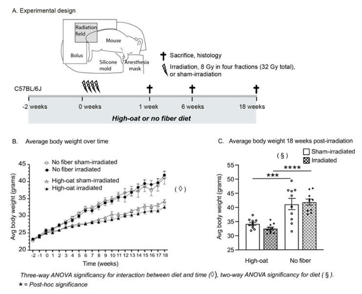
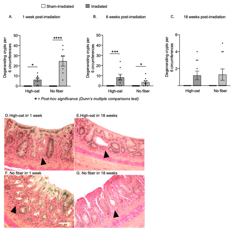
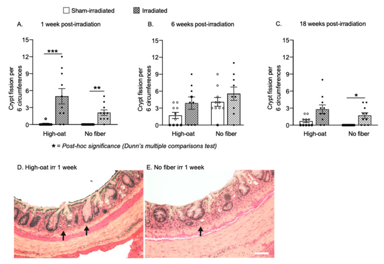
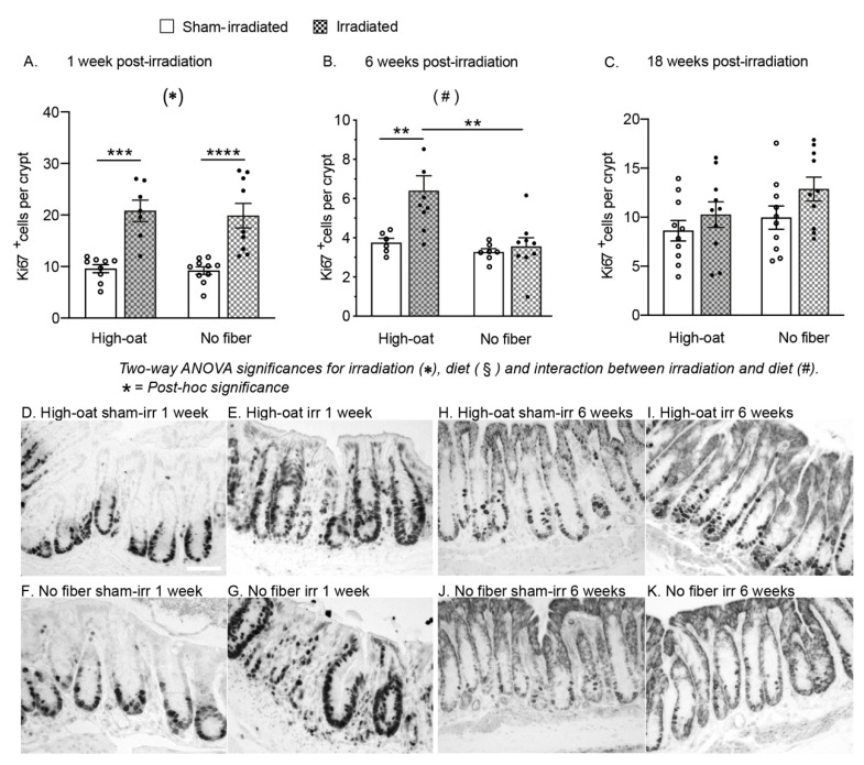
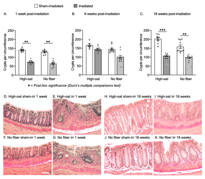
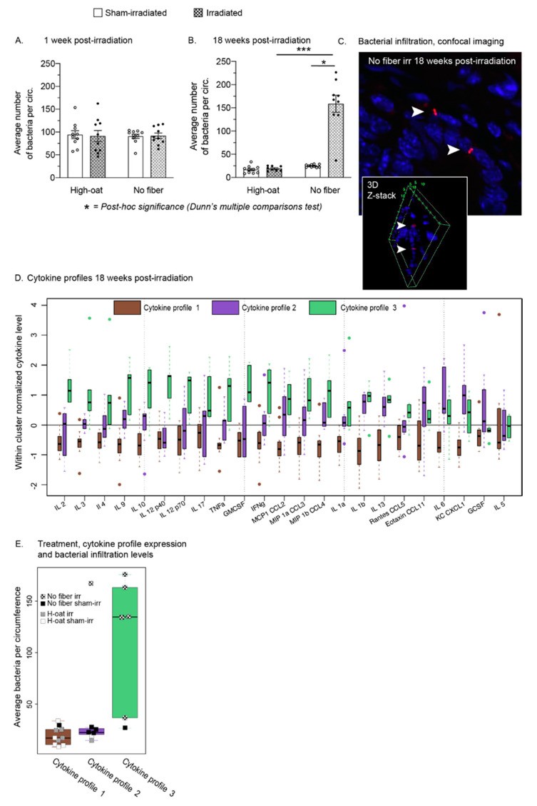
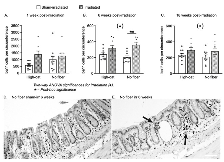
Dietary fiber is considered a strong intestinal protector, but we do not know whether dietary fiber protects against the long-lasting mucosal damage caused by ionizing radiation. To evaluate whether a fiber-rich diet can ameliorate the long-lasting pathophysiological hallmarks of the irradiated mucosa, C57BL/6J mice on a fiber-rich bioprocessed oat bran diet or a fiber-free diet received 32 Gray in four fractions to the distal colorectum using a linear accelerator and continued on the diets for one, six or 18 weeks. We quantified degenerating crypts, crypt fission, cell proliferation, crypt survival, macrophage density and bacterial infiltration. Crypt loss through crypt degeneration only occurred in the irradiated mice. Initially, it was most frequent in the fiber-deprived group but declined to levels similar to the fiber-consuming group by 18 weeks. The fiber-consuming group had a fast response to irradiation, with crypt fission for growth or healing peaking already at one week post-irradiation, while crypt fission in the fiber-deprived group peaked at six weeks. A fiber-rich diet allowed for a more intense crypt cell proliferation, but the recovery of crypts was eventually lost by 18 weeks. Bacterial infiltration was a late phenomenon, evident in the fiber-deprived animals and intensified manyfold after irradiation. Bacterial infiltration also coincided with a specific pro-inflammatory serum cytokine profile. In contrast, mice on a fiber-rich diet were completely protected from irradiation-induced bacterial infiltration and exhibited a similar serum cytokine profile as sham-irradiated mice on a fiber-rich diet. Our findings provide ample evidence that dietary fiber consumption modifies the onset, timing and intensity of radiation-induced pathophysiological processes in the intestinal mucosa. However, we need more knowledge, not least from clinical studies, before this finding can be introduced to a new and refined clinical practice.

Keywords: bacteria; colon; cytokines; dietary fiber; intestine; irradiation; oat.


Conflict of interest statement

A. Rascón is a part-time employee of Glucanova, the provider of the bioprocessed oat bran. She is also the author of the patent for its preparation.

Figures

Figure 1
Figure 1
(A). Experimental design: 9 week old male C57BL/6J mice were fed either a fiber-rich bioprocessed oat bran diet (“High-oat”) or a fiber-deprived diet (“No fiber”) for two weeks before irradiation or sham-irradiation. During irradiation, 32 Gray was given to the colorectum with a 6 MV linear accelerator, in four fractions of 8 Gy spaced by 12 h. The diets were continued throughout the experiment, and mice were euthanized 1 week (acute time point), 6 weeks (intermediate time point) or 18 weeks (late time point) after irradiation. The colorectal tissues were harvested for the collection of various metrics on injury and repair. (B). Weight curves of mice during the 20 weeks of the dietary intervention and up to 18 weeks after irradiation. Fiber-deprived mice gained more weight over time than mice fed with bioprocessed oat bran (p < 0.0001 for interaction between time and diet). (C). At 18 weeks after irradiation, mice on a fiber-free diet had gained significantly more weight than mice on a fiber-rich diet, and irradiation did not affect their weight gain. *** p < 0.0006, **** p < 0.0001. Data are shown as mean ± S.E.M.
Figure 2
Figure 2
Number of degenerating crypts at 1 week, 6 weeks and 18 weeks after irradiation. Degenerating crypts were never seen in sham-irradiated animals. (A). More crypt degeneration was seen in the fiber-deprived animals (“No fiber”) than the oat-fed animals (“High-oat”) at one week after irradiation. * p = 0.0191 and **** p < 0.0001. (B). Although the initial high rate of crypt degeneration in the irradiated No-fiber mice had declined by 6 weeks post-irradiation, crypt degeneration still occurred in both diet groups. *** p = 0.0008 and * p = 0.0205. (C). At 18 weeks, the High-oat and No-fiber irradiated mice both had similar and low numbers of degenerating crypts. (DG). Micrographs (40× magnification) of colorectal mucosa stained with Verhoeff’s Elastic Stain. Arrowheads depict examples of degenerating crypts. Scale bar 50 μm. Data are shown as mean ± S.E.M.
Figure 3
Figure 3
Number of crypt fissions per six circumferences at 1 week, 6 weeks and 18 weeks after irradiation. (A). An increase in the number of crypt fissions was observed in both the diet groups one week after irradiation, with a more pronounced increase in the High-oat group. *** p = 0.001 and ** p = 0.0034. (B). At six weeks, there was slight, but not statistically significant increase in the number of crypt fissions in both irradiated diet groups, and more crypt fissions in the animals fed the No-fiber diet. Both control groups had a higher baseline than the control groups at 1 and 18 weeks. (C). At 18 weeks, there were still more crypt fissions in the irradiated mice of both diet groups compared to the sham-irradiated mice. * p = 0.0157. (D,E). Micrographs (40× magnification) of colorectal mucosa stained with Verhoeff’s Elastic Stain. Arrows depict examples of crypt fissions. Data are shown as mean ± S.E.M.
Figure 4
Figure 4
The average number of Ki67-positive proliferating cells per mucosal crypt in mice fed a fiber-rich or a fiber-free diet. (A). An irradiation-induced increase in the number of Ki67-positive cells was found in both the High-oat and No-fiber animals at one week after irradiation. *** p = 0.0002, **** p = 0.0001. (B). At six weeks, an increase in the Ki67-positive cells was still observed in the High-oat irradiated mice but not in the No-fiber mice. ** p = 0.0088, ** p = 0.0015. (C). At 18 weeks, there was a slight increase in crypt cell proliferation in both irradiated groups, although this was not statistically significant. (DK). Micrographs (40× magnification) of proliferating cells visualized with immunohistochemistry using an antibody against Ki67 and a DAB stain. Scale bar 50 μm. Data are shown as mean ± S.E.M.
Figure 5
Figure 5
Crypts per circumference over time after irradiation. (A). A decrease in the number of crypts per circumference was seen in the High-oat and the No-fiber mice compared to the sham-irradiated mice at one week. ** p = 0.0025 and ** p = 0.0017. (B). At six weeks, the High-oat irradiated animals appeared to have more crypts compared to the No-fiber irradiated mice (p = 0.0771). (C). At 18 weeks, both the High-oat and the No-fiber irradiated animals had similar numbers of crypts. *** p = 0.0008 and ** p = 0.0025. (DK). Representative micrographs (40× magnification, Verhoeff’s Elastic Stain) of crypts in the colorectal mucosa. Data are shown as mean ± S.E.M.
Figure 6
Figure 6
The average number of bacteria per circumference at 1 week and 18 weeks after irradiation. (A). Quantification of bacteria in the mucosa one week after irradiation confirmed that bacterial infiltration did not occur early after irradiation, and the absence of dietary fiber in the chow did not yet facilitate infiltration. (B). At 18 weeks after irradiation, a manyfold increase of bacterial presence in the mucosa was found in irradiated mice on a fiber-free diet. Irradiated mice consuming fiber-rich oat bran were entirely protected against irradiation-induced bacterial infiltration. * p = 0.0451 and *** p = 0.0005. (C). Confocal imaging of bacteria (arrowhead; bacteria in red, cell nuclei in blue) in the irradiated colorectum of a mouse fed a fiber-free diet. Z-stack imaging shows the location of bacteria within the tissue. Data are shown as mean ± S.E.M. (D). Boxplots of within-cluster normalized serum cytokine levels at 18 weeks post-irradiation. The mice expressed either one of three distinct cytokine profiles. (E). The majority of irradiated mice on a fiber-free diet, with higher levels of bacterial infiltration, were clustering into profile 3, with a more pro-inflammatory pattern than profile 1 and 2. In contrast, mice on a fiber-rich diet expressed profile 1, regardless of whether they had been irradiated or not. Boxplots with median and interquartile range.
Figure 7
Figure 7
The average number of mucosal macrophages per circumference at 1 week, 6 weeks and 18 weeks after irradiation. (A). A potential increase in the number of macrophages was seen at one week in the High-oat irradiated mice compared to the High-oat sham-irradiated. (B). At 6 weeks, both diet groups had a greater abundancy of macrophages after irradiation than the sham-irradiated mice (p = 0.0002 for irradiation). ** p = 0.0022. (C). At 18 weeks, there were still more macrophages in the mucosa of irradiated mice of both diet groups (p = 0.0386 for irradiation). (D,E). Micrographs (20× magnification, scale bar 50 μm) of macrophages visualized with immunohistochemistry using an antibody against Iba1 and a DAB stain. Iba1+ macrophages were found in the mucosa of both sham-irradiated (D) and irradiated (E) animals but were more abundant in the irradiated animals and often clustered (arrows). Data are shown as mean ± S.E.M.

References

    1. Andreyev H.J., Wotherspoon A., Denham J.W., Hauer-Jensen M. “Pelvic radiation disease”: New understanding and new solutions for a new disease in the era of cancer survivorship. Scand. J. Gastroenterol. 2011;46:389–397. doi: 10.3109/00365521.2010.545832. - DOI - PubMed
    1. Steineck G., Skokic V., Sjöberg F., Bull C., Alevronta E., Dunberger G., Bergmark K., Wilderäng U., Oh J.H., Deasy J.O., et al. Identifying radiation-induced survivorship syndromes affecting bowel health in a cohort of gynecological cancer survivors. PLoS ONE. 2017;12:e0171461. doi: 10.1371/journal.pone.0171461. - DOI - PMC - PubMed
    1. Hur W., Yoon S.K. Molecular Pathogenesis of Radiation-Induced Cell Toxicity in Stem Cells. Int. J. Mol. Sci. 2017;18:2749. doi: 10.3390/ijms18122749. - DOI - PMC - PubMed
    1. Potten C.S., Grant H.K. The relationship between ionizing radiation-induced apoptosis and stem cells in the small and large intestine. Br. J. Cancer. 1998;78:993–1003. doi: 10.1038/bjc.1998.618. - DOI - PMC - PubMed
    1. Francois A., Milliat F., Guipaud O., Benderitter M. Inflammation and immunity in radiation damage to the gut mucosa. BioMed Res. Int. 2013;2013:123241. doi: 10.1155/2013/123241. - DOI - PMC - PubMed