. 2021 Dec 31;23(1):448.
doi: 10.3390/ijms23010448.

TGF-β1 Potentiates the Cytotoxicity of Cadmium by Induction of a Metal Transporter, ZIP8, Mediated by the ALK5-Smad2/3 and ALK5-Smad3-p38 MAPK Signal Pathways in Cultured Vascular Endothelial Cells

Affiliations

TGF-β1 Potentiates the Cytotoxicity of Cadmium by Induction of a Metal Transporter, ZIP8, Mediated by the ALK5-Smad2/3 and ALK5-Smad3-p38 MAPK Signal Pathways in Cultured Vascular Endothelial Cells

Keisuke Ito et al. Int J Mol Sci. .

Abstract

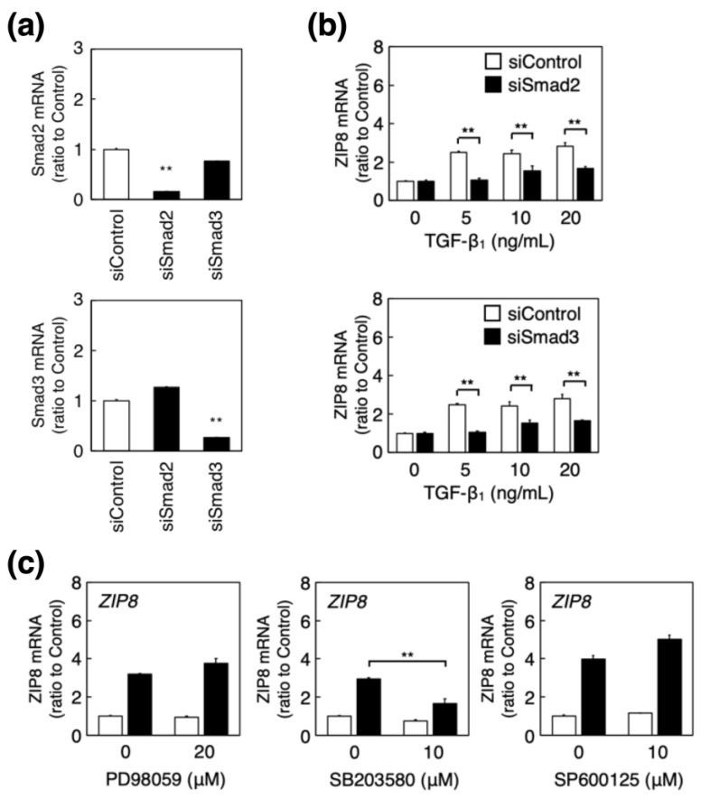
Vascular endothelial cells cover the luminal surface of blood vessels in a monolayer and play a role in the regulation of vascular functions, such as the blood coagulation-fibrinolytic system. When the monolayer is severely or repeatedly injured, platelets aggregate at the damaged site and release transforming growth factor (TGF)-β1 in large quantities from their α-granules. Cadmium is a heavy metal that is toxic to various organs, including the kidneys, bones, liver, and blood vessels. Our previous study showed that the expression level of Zrt/Irt-related protein 8 (ZIP8), a metal transporter that transports cadmium from the extracellular fluid into the cytosol, is a crucial factor in determining the sensitivity of vascular endothelial cells to cadmium cytotoxicity. In the present study, TGF-β1 was discovered to potentiate cadmium-induced cytotoxicity by increasing the intracellular accumulation of cadmium in cells. Additionally, TGF-β1 induced the expression of ZIP8 via the activin receptor-like kinase 5-Smad2/3 signaling pathways; Smad3-mediated induction of ZIP8 was associated with or without p38 mitogen-activated protein kinase (MAPK). These results suggest that the cytotoxicity of cadmium to vascular endothelial cells increases when damaged endothelial monolayers that are highly exposed to TGF-β1 are repaired.

Keywords: Smad2/3; Zrt- and Irt-like protein transporter; cadmium; endothelial cell; p38 MAPK; transforming growth factor-β1.


Conflict of interest statement

The authors declare no conflict of interest.

Figures

Figure 1
Figure 1
Influence of TGF-β1 on cadmium-induced cytotoxicity of vascular endothelial cells. Bovine aortic endothelial cells that were treated with or without TGF-β1 (1, 5, or 10 ng/mL) for 24 h were incubated in the presence or absence (white bars) of cadmium at 2 μM (thin gray bars) or 5 μM (dark gray bars) for 24 h. (a) This panel shows the cell layer stained with Giemsa. Original magnification (×40). Scale bar = 100 μm. (b) This panel shows the conditioned media used to determine lactate dehydrogenase activity, and (c) the intracellular accumulation of cadmium, which was measured using inductively coupled plasma-mass spectrometry. Each value represents the mean ± standard error (S.E.) of four independent experiments; and statistical significance compared to the corresponding cadmium-exposed cells without TGF-β1 was set as * p < 0.05, ** p < 0.01.
Figure 2
Figure 2
ZIP8 induction in vascular endothelial cells following treatment with TGF-β1. (a) Bovine aortic endothelial cells were treated with or without TGF-β1 (5, 10, or 20 ng/mL) for 24 h, and the expression of ZIP8 protein in the membrane fraction was determined using Western blotting. The band intensity of ZIP8 was normalized by coomassie brilliant blue (CBB) staining, and each value represents the mean ± S.E. of three independent samples; * p < 0.05, ** p < 0.01 compared with each control. (b) Bovine aortic endothelial cells were treated with or without TGF-β1 (1, 2, 5, 10 or 20 ng/mL) for 6 h (upper panel) and with (black circles) or without (white circles) TGF-β1 (10 ng/mL each) for 3, 6, 12 or 24 h (lower panel). ZIP8 mRNA levels were determined using real-time reverse transcription polymerase chain reaction (RT-PCR). Each value represents the mean ± S.E. of three technical replicates and ** p < 0.01 compared with the corresponding control. (c) Bovine aortic endothelial cells were treated with or without TGF-β1 (10 ng/mL) for 6 h, and ZIP1-4, ZIP6, ZIP7-14, ZnT1, and DMT1 mRNA expression levels were determined by real-time RT-PCR. Each value represents the mean ± S.E. of three technical replicates; ** p < 0.01 compared with the control.
Figure 3
Figure 3
Involvement of ALKs in the induction of ZIP8 by TGF-β1 in vascular endothelial cells. (a) Bovine aortic endothelial cells were transfected with control, ALK1, or ALK5 siRNAs for 24 h, and ALK1 and ALK5 mRNA expression levels were determined using real-time reverse transcription polymerase chain reaction (RT-PCR). Each value represents the mean ± S.E. of three technical replicates; and ** p < 0.01 represents statistical significance compared with the siControl. (b) Bovine aortic endothelial cells transfected with the control, ALK1, or ALK5 siRNA for 24 h were treated with or without TGF-β1 (5, 10, or 20 ng/mL) for 6 h, and ZIP8 mRNA expression was measured using real-time RT-PCR. Each value represents the mean ± S.E. of three technical replicates; * p < 0.05, ** p < 0.01 compared with the corresponding TGF-β1-treated cells transfected with siControl.
Figure 4
Figure 4
Involvement of Smad2/3 and MAPK signaling in the induction of ZIP8 by TGF-β1 in vascular endothelial cells. (a) Bovine aortic endothelial cells were transfected with control, Smad2, or Smad3 siRNAs for 24 h, and Smad2 and Smad3 mRNA expression levels were determined using real-time reverse transcription polymerase chain reaction (RT-PCR). Each value represents the mean ± S.E. of three technical replicates; ** p < 0.01 represents statistical significance compared with the siControl. (b) Bovine aortic endothelial cells transfected with control, Smad2, or Smad3 siRNAs for 24 h were treated with or without TGF-β1 (5, 10, or 20 ng/mL) for 6 h, and ZIP8 mRNA expression was determined using real-time RT-PCR. Each value represents the mean ± S.E. of three technical replicates; ** p < 0.01 compared with the corresponding siControl. (c) Bovine aortic endothelial cells treated with or without PD98059 (20 μM), SB203580, or SP600125 (10 μM each) for 3 h were treated with (black bars) or without (white bars) TGF-β1 at 5 ng/mL for 6 h, and ZIP8 mRNA expression levels were determined using real-time RT-PCR. Each value represents the mean ± S.E. of three technical replicates; ** p < 0.01 compared with the TGF-β1-treated cells without each inhibitor.
Figure 5
Figure 5
Interaction between TGF-β1-activated Smad2/3 and p38 MAPK signaling in vascular endothelial cells. (a) Bovine aortic endothelial cells treated with or without LY364947 (5 μM) for 24 h were treated with or without TGF-β1 (1, 2, 5, 10, or 20 ng/mL) for 6 h, and the expression levels of phosphorylated p38 MAPK (p-p38 MAPK) and total p38 MAPK proteins were determined using Western blotting. (b) Bovine aortic endothelial cells transfected with control, Smad2, or Smad3 siRNAs for 24 h were treated with or without TGF-β1 (5 or 20 ng/mL) for 6 h, and the expression levels of phosphorylated p38 MAPK (p-p38 MAPK) and total p38 MAPK proteins were determined using Western blotting. (c) Bovine aortic endothelial cells treated with or without SB203580 (10 μM) for 3 h that were treated with or without TGF-β1 (5 or 20 ng/mL) for 1 h, and the expression levels of phosphorylated Smad2/3 (p-Smad2/3) and total Smad2/3 proteins were determined using Western blotting.

Similar articles

Cited by

References

    1. Palmer R.M., Ferrige A.G., Moncada S. Nitric oxide release accounts for the biological activity of endothelium-derived relaxing factor. Nature. 1987;327:524–526. doi: 10.1038/327524a0. - DOI - PubMed
    1. Weksler B.B., Marcus A.J., Jaffe E.A. Synthesis of prostaglandin I2 (prostacyclin) by cultured human and bovine endothelial cells. Proc. Natl. Acad. Sci. USA. 1977;74:3922–3926. doi: 10.1073/pnas.74.9.3922. - DOI - PMC - PubMed
    1. Yanagisawa M., Kurihara H., Kimura S., Tomobe Y., Kobayashi M., Mitsui Y., Yazaki Y., Goto K., Masaki T. A novel potent vasoconstrictor peptide produced by vascular endothelial cells. Nature. 1988;332:411–415. doi: 10.1038/332411a0. - DOI - PubMed
    1. Levin E.G., Louskutoff D.J. Cultured bovine endothelial cells produce both urokinase and tissue-type plasminogen activators. J. Cell Biol. 1982;94:631–636. doi: 10.1083/jcb.94.3.631. - DOI - PMC - PubMed
    1. Gelehrter T.D., Sznycer-Laszuk R. Thrombin induction of plasminogen activator-inhibitor in cultured human endothelial cells. J. Clin. Investig. 1986;77:165–169. doi: 10.1172/JCI112271. - DOI - PMC - PubMed

MeSH terms

Substances

LinkOut - more resources