. 2021 Dec 31;23(1):451.
doi: 10.3390/ijms23010451.

Pulsed Electric Fields Alter Expression of NF-κB Promoter-Controlled Gene

Affiliations

Pulsed Electric Fields Alter Expression of NF-κB Promoter-Controlled Gene

Justina Kavaliauskaitė et al. Int J Mol Sci. .

Abstract

The possibility to artificially adjust and fine-tune gene expression is one of the key milestones in bioengineering, synthetic biology, and advanced medicine. Since the effects of proteins or other transgene products depend on the dosage, controlled gene expression is required for any applications, where even slight fluctuations of the transgene product impact its function or other critical cell parameters. In this context, physical techniques demonstrate optimistic perspectives, and pulsed electric field technology is a potential candidate for a noninvasive, biophysical gene regulator, exploiting an easily adjustable pulse generating device. We exposed mammalian cells, transfected with a NF-κB pathway-controlled transcription system, to a range of microsecond-duration pulsed electric field parameters. To prevent toxicity, we used protocols that would generate relatively mild physical stimulation. The present study, for the first time, proves the principle that microsecond-duration pulsed electric fields can alter single-gene expression in plasmid context in mammalian cells without significant damage to cell integrity or viability. Gene expression might be upregulated or downregulated depending on the cell line and parameters applied. This noninvasive, ligand-, cofactor-, nanoparticle-free approach enables easily controlled direct electrostimulation of the construct carrying the gene of interest; the discovery may contribute towards the path of simplification of the complexity of physical systems in gene regulation and create further synergies between electronics, synthetic biology, and medicine.

Keywords: NF-κB; cell line; inducible gene transcription control; mammalian cells; microsecond pulsed electric field; reporter assay; secreted alkaline phosphatase.


Conflict of interest statement

The authors declare no conflict of interest.

Figures

Figure 1
Figure 1
NF-κB/SEAP response system performance in different mammalian cell lines after UV irradiation. Indicated cell lines were transfected with a NF-κB-driven SEAP expression plasmid (pNF-κB-SEAP) and exposed to low (4.14 J/cm2) or high (20.7 J/cm2) UV doses 24 h before SEAP expression was quantified in the culture supernatant. Dashed line denotes baseline of negative control (cells cultivated under otherwise identical conditions, but in the absence of UV). Significances compared with control were determined using student’s t-test: * p < 0.05, ** p < 0.01.
Figure 2
Figure 2
(a) Membrane permeabilization after microsecond-duration pulsed electric fields treatment at the field strengths of 0.165, 0.25, 0.3, 0.4, 0.5, and 0.6 kV/cm. Eight square wave pulses of 100 µs-duration and 1 Hz-frequency were applied. SYTOX®-green stain uptake was measured one hour after pulsed electric field treatment. The dashed line denotes baseline of negative control (cells cultivated under otherwise identical conditions, but in the absence of pulsed electric fields). (b) Membrane reseal assay at E = 0.4 kV/cm. SYTOX®-green stain was added 0, 10, 30, 60, and 300 s after pulsed electric field treatment; fluorescence was read one hour later. Result significances compared with control were determined using student’s t-test: * p < 0.05, ** p < 0.01, *** p < 0.001, **** p < 0.0001.
Figure 3
Figure 3
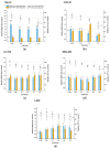
NF-κB/SEAP response system performance and viability readout in Hep-2c (a), CHO-K1 (b), U-2 OS (c), HEK-293 (d), and L-929 (e) cell lines after exposure to microsecond-duration pulsed electric fields. Hep-2c and CHO-K1 cells were stimulated with the electric field amplitudes of 0.165, 0.25, 0.3, and 0.4 kV/cm, eight pulses of 100 µs, frequency—1 Hz. U-2 OS, HEK-293, and L-929 cells were additionally simulated by applying the electric field amplitudes of 0.5 and 0.6 kV/cm. Results were read 24- and 48-h postexposure. Dashed lines denote baselines of negative SEAP and viability controls (cells cultivated under otherwise identical conditions, but in the absence of microsecond pulsed electric fields). Significances compared with control were determined using Kruskal–Wallis and Conover post hoc tests: * p < 0.05, ** p < 0.01, *** p < 0.001, **** p < 0.0001.
Figure 4
Figure 4
Hypothetical schematic representation of electrically modulated transcription of the SEAP reporter gene. Pulsed electric fields activate the nuclear factor κB pathway, leading to its release from the inhibitor and translocation to the nucleus. NF-κB binds to a synthetic promoter and activates the transcription of the SEAP reporter gene. The SEAP protein secretes to the cell growth medium and converts the substrate (p-nitrophenyl phosphate, PNPP) into a yellow detectable product.

Similar articles

Cited by

References

    1. Stuible M., Burlacu A., Perret S., Brochu D., Paul-Roc B., Baardsnes J., Loignon M., Grazzini E., Durocher Y. Optimization of a high-cell-density polyethylenimine transfection method for rapid protein production in CHO-EBNA1 cells. J. Biotechnol. 2018;281:39–47. doi: 10.1016/j.jbiotec.2018.06.307. - DOI - PubMed
    1. Lillacci G., Benenson Y., Khammash M. Synthetic control systems for high performance gene expression in mammalian cells. Nucleic Acids Res. 2018;46:9855–9863. doi: 10.1093/nar/gky795. - DOI - PMC - PubMed
    1. Rao R.C., Zacks D.N. Cell and Gene Therapy. Cell-Based Ther. Retin. Degener. Dis. 2014;53:167–177. doi: 10.1159/000357376. - DOI - PMC - PubMed
    1. Lienert F., Lohmueller J., Garg A.D., Silver P.A. Synthetic biology in mammalian cells: Next generation research tools and therapeutics. Nat. Rev. Mol. Cell Biol. 2014;15:95–107. doi: 10.1038/nrm3738. - DOI - PMC - PubMed
    1. Re A. Synthetic Gene Expression Circuits for Designing Precision Tools in Oncology. Front. Cell Dev. Biol. 2017;5:77. doi: 10.3389/fcell.2017.00077. - DOI - PMC - PubMed

Grants and funding

LinkOut - more resources