Review
. 2021 Dec 31;23(1):452.
doi: 10.3390/ijms23010452.

Microvascular Experimentation in the Chick Chorioallantoic Membrane as a Model for Screening Angiogenic Agents including from Gene-Modified Cells

Affiliations
Review

Microvascular Experimentation in the Chick Chorioallantoic Membrane as a Model for Screening Angiogenic Agents including from Gene-Modified Cells

Donna C Kennedy et al. Int J Mol Sci. .

Abstract

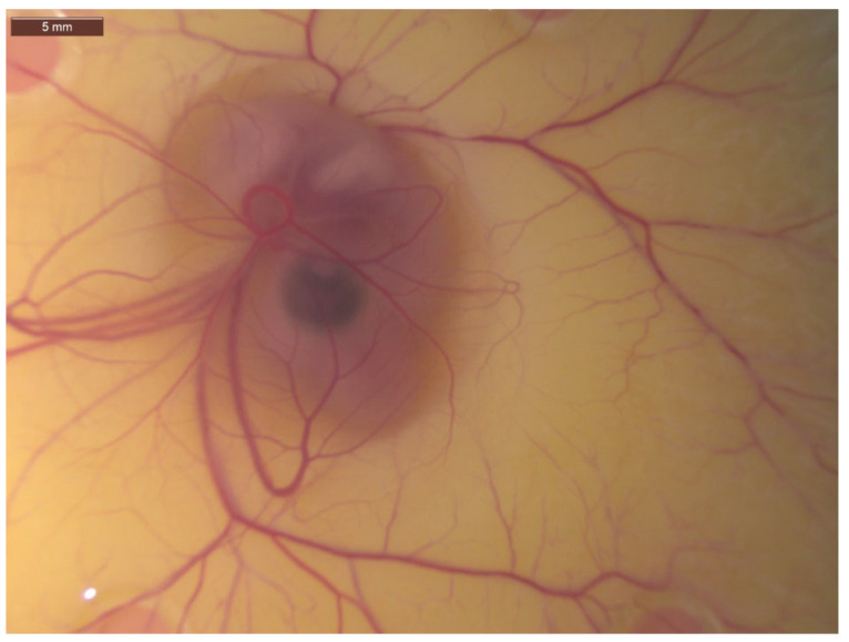
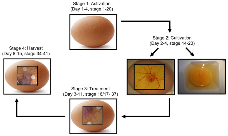
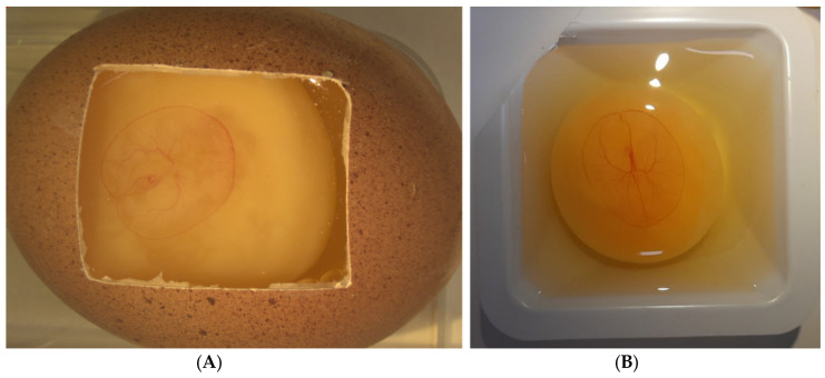
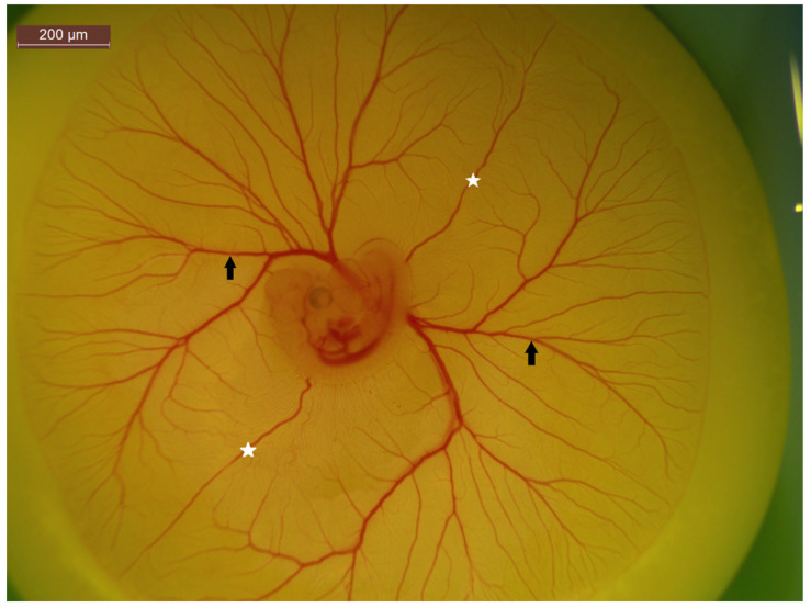
The chick chorioallantoic membrane (CAM) assay model of angiogenesis has been highlighted as a relatively quick, low cost and effective model for the study of pro-angiogenic and anti-angiogenic factors. The chick CAM is a highly vascularised extraembryonic membrane which functions for gas exchange, nutrient exchange and waste removal for the growing chick embryo. It is beneficial as it can function as a treatment screening tool, which bridges the gap between cell based in vitro studies and in vivo animal experimentation. In this review, we explore the benefits and drawbacks of the CAM assay to study microcirculation, by the investigation of each distinct stage of the CAM assay procedure, including cultivation techniques, treatment applications and methods of determining an angiogenic response using this assay. We detail the angiogenic effect of treatments, including drugs, metabolites, genes and cells used in conjunction with the CAM assay, while also highlighting the testing of genetically modified cells. We also present a detailed exploration of the advantages and limitations of different CAM analysis techniques, including visual assessment, histological and molecular analysis along with vascular casting methods and live blood flow observations.

Keywords: angiogenesis; blood flow; cancer; chorioallantoic membrane (CAM); microcirculation; tumour.


Conflict of interest statement

The authors declare no conflict of interest.

Figures

Figure 1
Figure 1
Image of 7-day-old chick embryo with associated chick chorioallantoic membrane (CAM) and its vast vascular network of capillaries, veins and arteries visible. Image taken at 25× magnification.
Figure 2
Figure 2
Schematic of four-stage CAM assay process, along with approximate embryonic development days where this stage typically takes place. Stage 1: Activation is where eggs are put in a rotating incubator at 50% humidity to allow for preliminary development. Stage 2: Cultivation allows for visualisation of the embryo and CAM through either ex ovo cultivation where the eggshell is cracked with contents then transferred into a sterile petri dish, or in ovo cultivation where a saw tool is used to excise a window in the surface of the eggshell. Stage 3: Treatments such as cells, drugs or growth factors are applied. This can be through a variety of methods such as application of on-plants, pipetting directly onto the CAM surface or injection into the CAM vasculature. Finally, upon completion of the experiment, the chick embryo is sacrificed and the CAM is removed for analysis. Analysis can include visual observations of angiogenesis, histological examination, or molecular investigation.
Figure 3
Figure 3
4-day old chick embryo and associated chorioallantoic membrane (CAM) following (A) In ovo and (B) Ex ovo cultivation. The CAM expands as the embryo grows. Ex ovo cultivation is beneficial through the larger surface area available for experimentation, however embryo survival is impacted.
Figure 4
Figure 4
A 5-day-old chick embryo highlight CAM vasculature. The white stars represent anterior and posterior vitelline veins, while the black arrows indicate vitelline arteries and veins. The non-branching nature of the vitelline veins make it an ideal location for injections.
Figure 5
Figure 5
Examples of CAM analysis techniques to quantify angiogenic score following treatment. In the case of each of these methods, each blood vessel which fits specific criteria is given a score, with the accumulative score then determined for each on-plant/treatment. (A) Centripetal ordering method of angiogenic scoring, where vessels are assigned a score based on the order of their branching, with higher order vessels getting a higher score as described in DeFouw et al. [231]. (B) A range of concentric circles projected onto an image of a CAM where the total vascular index quantified based on the intersection of blood vessels with each of the circle, as described in Burggren et al. [230]. (C) Evaluation of a angiogenic response by scoring vessel branching as described by Ribatti et al. [32], this method involves the assigning of an angiogenic score ranging from 0–2 based on branching and angle of approach.

Similar articles

Cited by

References

    1. Giaccia A.J., Simon M.C., Johnson R. The biology of hypoxia: The role of oxygen sensing in development, normal function, and disease. Genes Dev. 2004;18:2183–2194. doi: 10.1101/gad.1243304. - DOI - PMC - PubMed
    1. Grace P. Ischaemia-reperfusion injury. Br. J. Surg. 1994;81:637–647. doi: 10.1002/bjs.1800810504. - DOI - PubMed
    1. AS M.N., Deshpande R., Kale V.P., Bhonde R.R., Datar S.P. Establishment of an in ovo chick embryo yolk sac membrane (YSM) assay for pilot screening of potential angiogenic and anti-angiogenic agents. Cell Biol. Int. 2018;42:1474–1483. doi: 10.1002/cbin.11051. - DOI - PubMed
    1. Ribatti D., Crivellato E. “Sprouting angiogenesis”, a reappraisal. Dev. Biol. 2012;372:157–165. doi: 10.1016/j.ydbio.2012.09.018. - DOI - PubMed
    1. Ausprunk D., Knighton D., Folkman J. Vascularization of normal and neoplastic tissues grafted to the chick chorioallantois. Role of host and preexisting graft blood vessels. Am. J. Pathol. 1975;79:597. - PMC - PubMed

MeSH terms

Substances

LinkOut - more resources