. 2021 Dec 31;23(1):462.
doi: 10.3390/ijms23010462.

Relevance of a Novel Circuit-Level Model of Episodic Memories to Alzheimer's Disease

Affiliations

Relevance of a Novel Circuit-Level Model of Episodic Memories to Alzheimer's Disease

Krisztián A Kovács. Int J Mol Sci. .

Abstract

The medial temporal lobe memory system has long been identified as the brain region showing the first histopathological changes in early Alzheimer's disease (AD), and the functional decline observed in patients also points to a loss of function in this brain area. Nonetheless, the exact identity of the neurons and networks that undergo deterioration has not been determined so far. A recent study has identified the entorhinal and hippocampal neural circuits responsible for encoding new episodic memories. Using this novel model we describe the elements of the episodic memory network that are especially vulnerable in early AD. We provide a hypothesis of how reduced reelin signaling within such a network can promote AD-related changes. Establishing novel associations and creating a temporal structure for new episodic memories are both affected in AD. Here, we furnish a reasonable explanation for both of these previous observations.

Keywords: Alzheimer’s disease; EPISODE module; anterograde amnesia; entorhinal cortex; episodic memories; granule cells; hippocampus; neurogenesis; reelin; synaptogenesis.


Conflict of interest statement

The author declares no conflict of interest.

Figures

Figure 1
Figure 1
The key actors of reelin signaling. The two neuronal receptors of reelin are depicted in green, along with their downstream effector molecules. On the left, we show the mechanism of how the cellular migration and the dendritogeneis are promoted by reelin, while on the right the main intracellular pathway leading to synaptic plasticity and synaptogenesis is highlighted. Both mechanisms are essential for the newborn granule cells to establish functional contacts with entorhinal input axons. More than two decades of research link AD to a deficit of adult reelin signaling hence some key reports describing prominent links to particular signaling molecules are indicated and coupled to those molecules with a red arrow.
Figure 2
Figure 2
Molecular, cellular, and network mechanisms of encoding new associations. The entities to be linked together in an episodic memory trace have previously encoded representations in the central nervous system that are relayed to the hippocampus by the ring attractors located in the EPISODE modules of the entorhinal cortex. Namely, entities A, B, C, and D are respectively represented by their own A, B, C, and D ring attractors which are activated by relevant sensory input, integrated input derived from multimodal external information, or internally generated input arriving actually to the entorhinal cortex. Axons from neurons (depicted in green, labeled as “FAN” cells) constituting the ring attractors reach the hippocampus via the perforant pathway. When encoding new episodic memories, new associations require newborn dentate granule cells (depicted as “GC”) that are of the right receptive age to form new synapses (indicated as red dots) with such axons. Reelin (see Figure 1) is indispensable for the newborn cells to become postsynaptic cells in this circuit and is provided by the output cells (fan cells or stellate cells) of the EPISODE module. New associations are stored at the level of the granule cells since these neurons establish contacts with a unique set (in the depicted example: A, B, C, and D) of EPISODE modules. For details, the reader is referred to Reference [3].
Figure 3
Figure 3
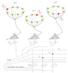
Molecular, cellular, and network mechanisms of encoding sequences. The internal temporal structure of any episodic memory is established by first assigning a theta phase (red inset diagram depicted next to the ring attractors) to each entity represented (three are shown in the Figure: A, B, and C) by a currently active EPISODE module. This theta phase serves to indicate whether the entity is upcoming, current, or already experienced and is repeated several times as several theta cycles unfold during the mnemonic episode to be encoded. As the temporal status of an entity (A, B, or C) changes, a directly measurable phenomenon, the so-called “phase precession” [59] is observed. Importantly, the generation and the repetition of the phase code in the entorhinal cortex is simply explained by the intrinsic electrophysiological and network properties of ring attractors. The theta phase is first transmitted to the dentate granule cells (depicted as “GC”) via the perforant pathway, then to the pyramidal CA3 neurons of the hippocampus via the mossy fibers as indicated by inset diagrams. Sequences are generated in the recurrent network of the CA3 using the phase code derived from the entorhinal EPISODE modules by the following mechanism: adjacent elements of the sequence produce coincident signals at the recurrent CA3-CA3 synapses given that the earlier signal (such as “A” in the Figure) suffers a delay when traveling along the recurrent collaterals and thus becomes coincident with a later one (such as “B” in the Figure) thereby making NMDA dependent plasticity possible (grey dot encircled with red). Sequences are generated by linking adjacent pairs first (in the Figure binding B to A and binding C to B is shown). The Figure is provided only to demonstrate the principle, therefore, several aspects are strongly simplified: (i) further cell types of the entorhinal ring attractors are not shown; (ii) the theta phase is also shifted during the entorhino–hippocampal transmission of the signal (inheritance of the phase precession), however, this results in the same amount of delay for all the entities (A, B, C) thus this aspect is not shown, and (iii) all the entities are represented by a population of cells, therefore, a population of “A” neurons becomes bound to a population of “B” neurons in CA3, etc.; however, for the sake of simplicity, only one neuron represents each population in the Figure. For details, the reader is referred to Reference [3].

Similar articles

Cited by

References

    1. Vogel J.W., Iturria-Medina Y., Strandberg O.T., Smith R., Levitis E., Evans A.C., Hansson O., Alzheimer’s Disease Neuroimaging Initiative. the Swedish BioFinder Study Spread of pathological tau proteins through communicating neurons in human Alzheimer’s disease. Nat. Commun. 2020;11:2612. doi: 10.1038/s41467-020-15701-2. - DOI - PMC - PubMed
    1. Minati L., Edginton T., Bruzzone M.G., Giaccone G. Reviews: Current Concepts in Alzheimer’s Disease: A Multidisciplinary Review. Am. J. Alzheimer’s Dis. Other Dement. 2009;24:95–121. doi: 10.1177/1533317508328602. - DOI - PMC - PubMed
    1. Kovács K.A. Episodic Memories: How do the Hippocampus and the Entorhinal Ring Attractors Cooperate to Create Them? Front. Syst. Neurosci. 2020;14:559168. doi: 10.3389/fnsys.2020.559186. - DOI - PMC - PubMed
    1. Talamonti D., Koscik R., Johnson S., Bruno D. Temporal contiguity and ageing: The role of memory organization in cognitive decline. J. Neuropsychol. 2021;15:53–65. doi: 10.1111/jnp.12219. - DOI - PMC - PubMed
    1. Witter M.P., Doan T.P., Jacobsen B., Nilssen E.S., Ohara S. Architecture of the Entorhinal Cortex A Review of Entorhinal Anatomy in Rodents with Some Comparative Notes. Front. Syst. Neurosci. 2017;11:46. doi: 10.3389/fnsys.2017.00046. - DOI - PMC - PubMed