. 2022 Jan 4;23(1):551.
doi: 10.3390/ijms23010551.

Thymosin β4 Is an Endogenous Iron Chelator and Molecular Switcher of Ferroptosis

Affiliations

Thymosin β4 Is an Endogenous Iron Chelator and Molecular Switcher of Ferroptosis

Joanna I Lachowicz et al. Int J Mol Sci. .

Abstract

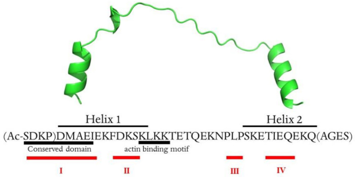
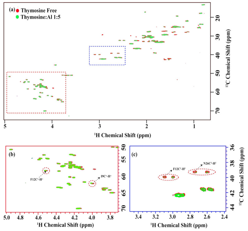
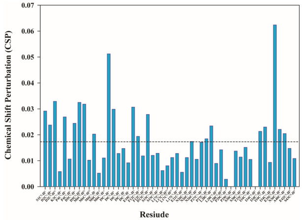
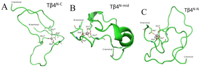
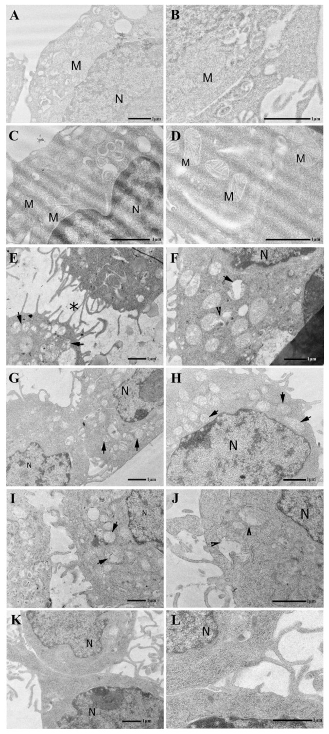
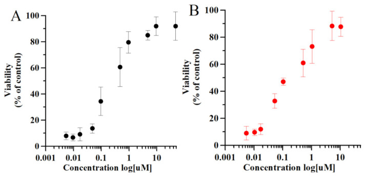
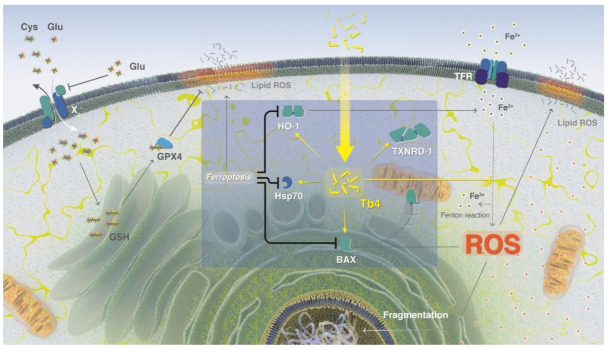
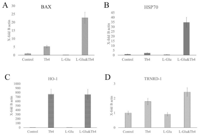
Thymosin β4 (Tβ4) was extracted forty years agofrom calf thymus. Since then, it has been identified as a G-actin binding protein involved in blood clotting, tissue regeneration, angiogenesis, and anti-inflammatory processes. Tβ4 has also been implicated in tumor metastasis and neurodegeneration. However, the precise roles and mechanism(s) of action of Tβ4 in these processes remain largely unknown, with the binding of the G-actin protein being insufficient to explain these multi-actions. Here we identify for the first time the important role of Tβ4 mechanism in ferroptosis, an iron-dependent form of cell death, which leads to neurodegeneration and somehow protects cancer cells against cell death. Specifically, we demonstrate four iron2+ and iron3+ binding regions along the peptide and show that the presence of Tβ4 in cell growing medium inhibits erastin and glutamate-induced ferroptosis in the macrophage cell line. Moreover, Tβ4 increases the expression of oxidative stress-related genes, namely BAX, hem oxygenase-1, heat shock protein 70 and thioredoxin reductase 1, which are downregulated during ferroptosis. We state the hypothesis that Tβ4 is an endogenous iron chelator and take part in iron homeostasis in the ferroptosis process. We discuss the literature data of parallel involvement of Tβ4 and ferroptosis in different human pathologies, mainly cancer and neurodegeneration. Our findings confronted with literature data show that controlled Tβ4 release could command on/off switching of ferroptosis and may provide novel therapeutic opportunities in cancer and tissue degeneration pathologies.

Keywords: NMR; TEM; ferroptosis; mRNA; metal chelation; molecular dynamics; thymosine beta 4.


Conflict of interest statement

The authors declare no conflict of interest.

Figures

Scheme 1
Scheme 1
Primary sequence and 3D structure of Tβ4 (PDB: 4PL7). Amino acids in parentheses were not defined by the X-ray structure and were added using Modeller v. 10.0 following the proper secondary structure elements (alpha helix). The metal-binding sites (I–IV) are signed in red. The scheme was created by Joanna Izabela Lachowicz, University of Cagliari. This image is original and designed specifically for the targeted publication. It should not be cropped, distorted, or in any way edited without the expressed consent of Research Publication Services.
Figure 1
Figure 1
(a) The overlay of the two-dimensional 13C-1H HSQC spectra in free form (red) and with thymosin: Al3+ ratio 1:5 (green). The alpha and beta proton regions are highlighted in the red and blue dotted rectangles in (a) and are presented in (b,c), respectively. Noticeable chemical shift changes were observed for F12, I9 as indicated by red circles in (b). Otherwise, the chemical shifts remain the same, with only some decrease in the intensities, as highlighted by the red circles in (c).
Figure 2
Figure 2
The chemical shift perturbation (CSP) plot for the 48 distinct peaks from the overlay of the two-dimensional 13C-1H HSQC spectra in free form and with thymosin:Al3+ ratio 1:5.The formula used for CSP is Sqrt [1/2(δH2 + α ∗ δ13C2)] as suggested by Willamson [26]. The α was kept 0.3.
Figure 3
Figure 3
Representative structures of (A) Tβ4N-C; (B) Tβ4N-mid and (C) Tβ4N-N. Figure was created by Gabriele dalla Torre, Euskal Herriko Unibertsitatea UPV/EHU. This image is original and designed specifically for the targeted publication. It should not be cropped, distorted, or in any way edited without the expressed consent of Research Publication Services.
Figure 4
Figure 4
Electron micrographs of J774cells. (A,B) The Control group displays a well-preserved ultrastructure. (C,D) Administration of Tβ4 (10 μM, 24 h) does not appear to alter cell morphology. Note in both control and Tβ4 treated samples the presence of several mitochondria showing a continuous mitochondria membrane and well-organized cristae. M = mitochondria, N = nucleus. (E,F) Glutamate treatment (5.5 mM, 24 h) induces numerous cellular alterations that affect especially the mitochondria (arrows). Initially, it appears that the membranes of the mitochondria open up, letting the contents pour out (arrowhead). Note the presence of tunneling nanotubes connecting adjacent cells (asterisk). (G,H) After Tβ4 administration (10 μM, 20 h), the cytoplasmic compartment displays more preserved cell structures. Mitochondria show a continuous mitochondrial membrane and partially reorganized cristae. N = nucleus. (I,J) Erastin treatment (0.5 μM; 24 h) induces numerous cellular alterations that affect especially the mitochondria (arrows). Mitochondria are frequently observed with ruptured mitochondrial membrane and altered cristae (arrowheads). (K,L) After Tβ4 administration (10 μM, 20 h), the cytoplasmic compartment displays more preserved cell structures associated with an evident decrease in lysosomal activity. N = nucleus.
Figure 5
Figure 5
Dose-response relationship for inhibition of erastin-induced (10 μM, 24 h) death in J774 cells by Fer-1 (A) and Tβ4 (B). Cell viability quantitative data are shown as mean and standard error. Statistical comparison of the cell viability quantitative data in different experimental conditions were performed, with 5.0 GraphPad Prism software, by the unpaired t-test. The data were also analyzed with one-way ANOVA. p values < 0.05 was considered significant.
Scheme 2
Scheme 2
Tβ4 inhibitory activity of ferroptosis. An excess of L-Glu in the extracellular matrix leads to the inhibition of GSH synthesis and reduced anti-oxidative activity, which increases the concentration of free iron ions in the cell and enhances the production of reactive oxygen species (ROS). High ROS and free iron ion concentrations trigger ferroptosis. Ferroptosis leads to lipid membrane disruption by ROS, and silences the expression of HO-1, Hsp70, and BAX. Extracellular treatment with Tβ4 can inhibit ferroptosis by increasing the expression of HO-1, Hsp70, BAX, and TXNRD-1, and direct chelation of free iron ions. The scheme was created by Heno Hwang, scientific illustrator at King Abdullah University of Science and Technology (KAUST). This image is original and designed specifically for the targeted publication. It should not be cropped, distorted, or in any way edited without the expressed consent of Research Publication Services.
Figure 6
Figure 6
mRNA expression levels (mean and standard error) of (A) BAX, (B) HSP70 (C) HO-1, and (D) TRNRD-1, which are related to stress conditions, in J774 cells. The amount of mRNA of each gene for each experiment was normalized with the respective value of the beta-actin housekeeping gene mRNA (X-fold of beta-actin expression). Statistical comparison of the gene’s expression in different experimental conditions was performed with 5.0 GraphPad Prism software. The data were also analyzed with one-way ANOVA. p values < 0.05 was considered significant.

Similar articles

Cited by

References

    1. Dixon S.J. Ferroptosis: Bug or feature? Immunol. Rev. 2017;277:150–157. doi: 10.1111/imr.12533. - DOI - PubMed
    1. Galluzzi L., Vitale I., Aaronson S.A., Abrams J.M., Adam D., Agostinis P., Alnemri E.S., Altucci L., Amelio I., Andrews D.W., et al. Molecular mechanisms of cell death: Recommendations of the Nomenclature Committee on Cell Death 2018. Cell Death Differ. 2018;25:486–541. doi: 10.1038/s41418-017-0012-4. - DOI - PMC - PubMed
    1. Dixon S.J., Lemberg K.M., Lamprecht M.R., Skouta R., Zaitsev E.M., Gleason C.E., Patel D.N., Bauer A.J., Cantley A.M., Yang W.S., et al. Ferroptosis: An Iron-Dependent Form of Nonapoptotic Cell Death. Cell. 2012;149:1060–1072. doi: 10.1016/j.cell.2012.03.042. - DOI - PMC - PubMed
    1. Yagoda N., von Rechenberg M., Zaganjor E., Bauer A.J., Yang W.S., Fridman D.J., Wolpaw A.J., Smukste I., Peltier J.M., Boniface J.J. RAS–RAF–MEK-dependent oxidative cell death involving voltage-dependent anion channels. Nature. 2007;447:865–869. doi: 10.1038/nature05859. - DOI - PMC - PubMed
    1. Ndayisaba A., Kaindlstorfer C., Wenning G.K. Iron in neurodegeneration–cause or consequence? Front. Neurosci. 2019;13:180. doi: 10.3389/fnins.2019.00180. - DOI - PMC - PubMed