. 2022 Jan 5;23(1):553.
doi: 10.3390/ijms23010553.

Indigo Pulverata Levis (Chung-Dae, Persicaria tinctoria) Alleviates Atopic Dermatitis-like Inflammatory Responses In Vivo and In Vitro

Affiliations

Indigo Pulverata Levis (Chung-Dae, Persicaria tinctoria) Alleviates Atopic Dermatitis-like Inflammatory Responses In Vivo and In Vitro

Ga-Yul Min et al. Int J Mol Sci. .

Abstract

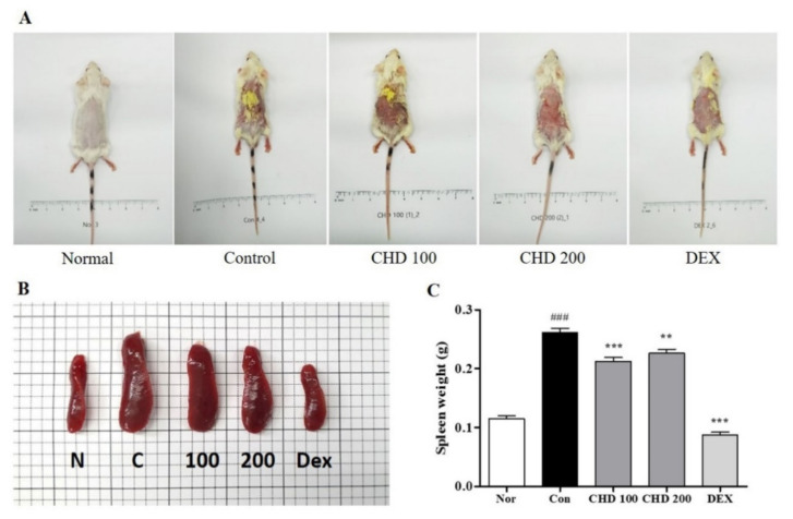
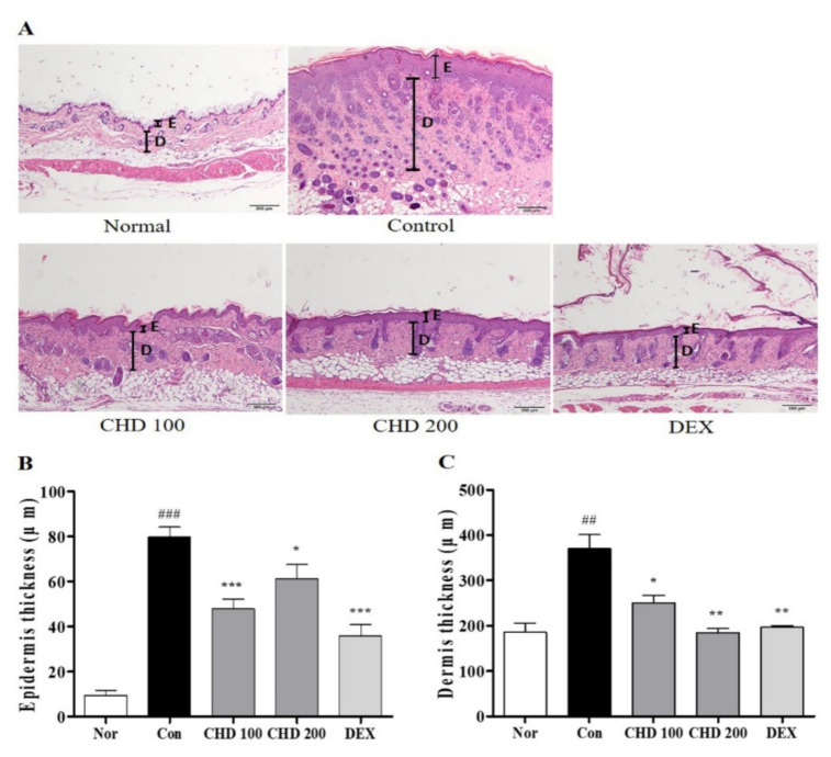
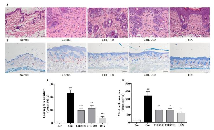
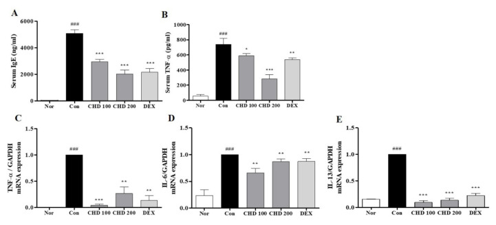
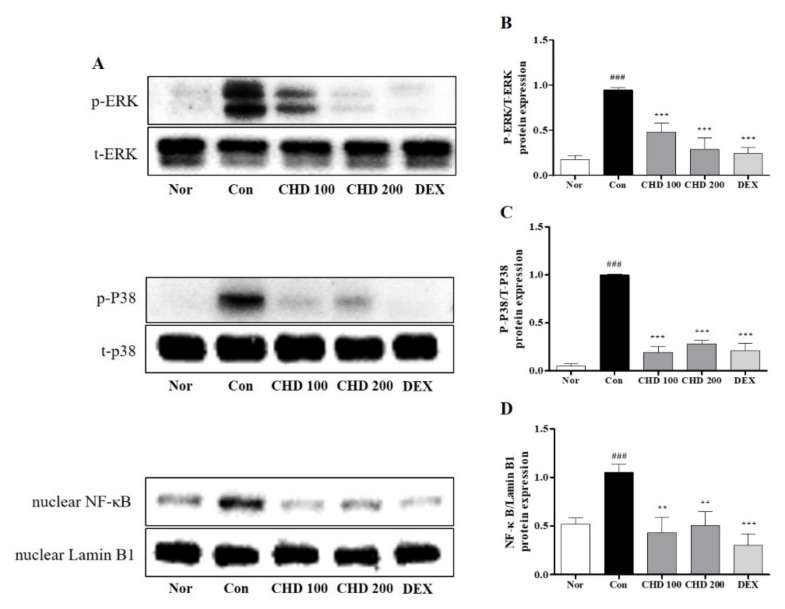
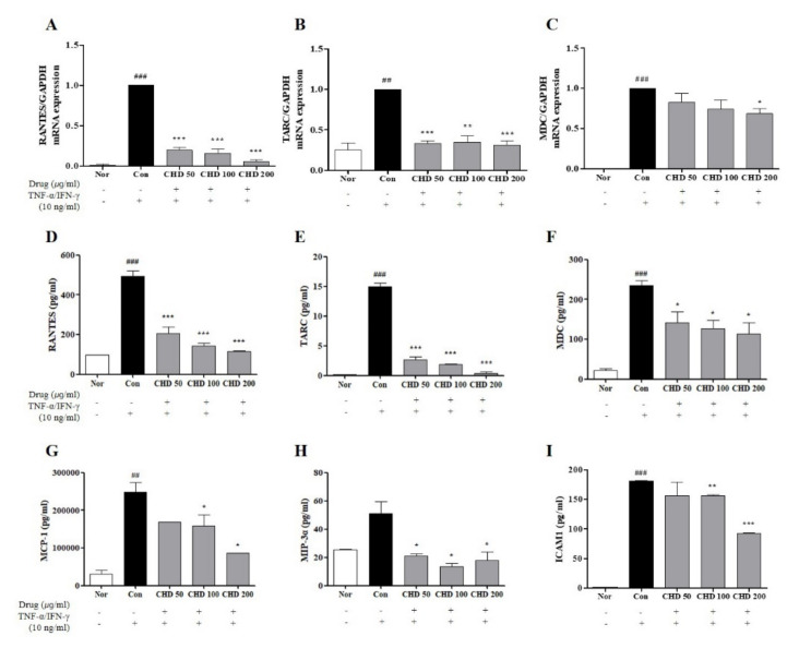
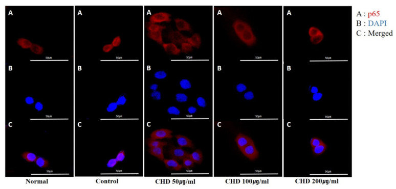
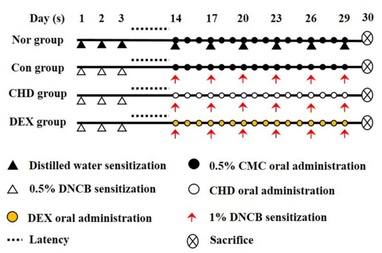
Atopic dermatitis (AD) is a chronic inflammatory skin disease associated with a type 2 T helper cell (Th2) immune response. The IndigoPulverata Levis extract (CHD) is used in traditional Southeast Asian medicine; however, its beneficial effects on AD remain uninvestigated. Therefore, we investigated the therapeutic effects of CHD in 2,4-dinitrochlorobenzene (DNCB)-induced BALB/c mice and tumor necrosis factor (TNF)-α- and interferon gamma (IFN)-γ-stimulated HaCaT cells. We evaluated immune cell infiltration, skin thickness, and the serum IgE and TNF-α levels in DNCB-induced AD mice. Moreover, we measured the expression levels of pro-inflammatory cytokines, mitogen-activated protein kinase (MAPK), and the nuclear factor-kappa B (NF-κB) in the mice dorsal skin. We also studied the effect of CHD on the translocation of NF-κB p65 and inflammatory chemokines in HaCaT cells. Our in vivo results revealed that CHD reduced the dermis and epidermis thicknesses and inhibited immune cell infiltration. Furthermore, it suppressed the proinflammatory cytokine expression and MAPK and NF-κB phosphorylations in the skin tissue and decreased serum IgE and TNF-α levels. In vitro results indicated that CHD downregulated inflammatory chemokines and blocked NF-κB p65 translocation. Thus, we deduced that CHD is a potential drug candidate for AD treatment.

Keywords: Indigo Pulverata Levis; NF-κB p65; atopic dermatitis; immune-cell infiltration; proinflammatory cytokines; skin thickness.


Conflict of interest statement

The authors declare no conflict of interest.

Figures

Figure 1
Figure 1
Effects of the Indigo Pelvarata Levis extract (CHD) on dorsal skin lesions and spleen hypertrophy in mice with atopic dermatitis (AD). (A,B) Photographs of dorsal skin lesions and spleens in AD mice. (C) Measurement of spleen hypertrophy. Data represent the mean ± standard error of the mean. n = 10. ### p < 0.001 vs. normal group; ** p < 0.01 and *** p < 0.001 vs. DNCB-induced group.
Figure 2
Figure 2
Effects of the Indigo Pelvarata Levis extract (CHD) on the histological characteristics of atopic dermatitis mice models. (A) Epidermis and dermis thicknesses were examined by H&E staining (100× magnification; scale bar: 200 µm). (B,C) Measurement of epidermis and dermis thicknesses. Data represent the mean ± standard error of the mean. n = 10. ## p < 0.001 and ### p < 0.01 vs. the normal group; * p < 0.05, ** p < 0.01, and *** p < 0.001 vs. the DNCB-induced control group.
Figure 3
Figure 3
Effects of the Indigo Pelvarata Levis extract (CHD) on immune cell infiltration in atopic dermatitis mice models. (A,B) H&E and toluidine blue stained eosinophils and mast cells that infiltrate into dermis lesions (H&E: 400× magnification, 50 µm scale bar; toluidine blue: 100× magnification, 200 µm scale bar). (C,D) The number of infiltrating immune cells quantified using the ImageJ software. Data represent the mean ± standard error of the mean. n = 10. ## p < 0.001 and ### p < 0.01 vs. the normal group; * p < 0.05, ** p < 0.01, and *** p < 0.001 vs. the DNCB-induced control group.
Figure 4
Figure 4
Effects of the Indigo Pelvarata Levis extract (CHD) on the IgE and pro-inflammatory cytokine levels in atopic dermatitis mice models. (A,B) The serum IgE and TNF-α levels were analyzed by the enzyme-linked immunosorbent assay. (CE) mRNA expression levels of the cytokines TNF-α, IL-6, and IL-13 were determined by real-time RT-PCR. Data represent the mean ± standard error of the mean. n = 10. ### p < 0.01 vs. the normal group; * p < 0.05, ** p < 0.01 and *** p < 0.001 vs. the DNCB-induced group.
Figure 5
Figure 5
Effects of the Indigo Pelvarata Levis extract (CHD) on the MAPK/NF-κB signaling pathway in atopic dermatitis mice models. (A) p-ERK, p-p38, and NF-κB expression levels analyzed by Western blotting. (BD) Evaluation of the MAPK and NF-κB expression levels using ImageJ. Data are represented as mean ± standard error of the mean. n = 5. ### p < 0.01 vs. the normal group; ** p < 0.01 and *** p < 0.001 vs. the DNCB-induced group.
Figure 6
Figure 6
Effects of the Indigo Pelvarata Levis extract (CHD) on the chemokine expression levels in HaCaT cells. (AC) Production levels of RANTES, TARC, and MDC were determined by real-time RT-PCR. (DI) Levels of RANTES, TARC, MDC, MCP-1, MIP-3α, and ICAM1 examined by ELISA. Data represent the mean ± standard error of the mean of three independent experiments. ## p < 0.001 and ### p < 0.01 vs. the normal group; * p < 0.05, ** p < 0.01, and *** p < 0.001 vs. the control group.
Figure 7
Figure 7
Effects of the Indigo Pelvarata Levis extract (CHD) on NF-κB p65 translocation in HaCaT cells. (AC) Translocation of p65 was analyzed using a fluorescent microscopy. Representative photomicrographs of (A) NF-κB p65 (red), (B) DAPI (blue), and (C) merged images (nuclear/cytosol) in HaCaT cells.
Figure 8
Figure 8
Representative chromatograms of the Indigo Pelvarata Levis extract (CHD). (A) Chemical structures of indigo and indirubin. (B) Standard indigo and indirubin solutions and the CHD extract.
Figure 9
Figure 9
Schematic representation of the study design.

Similar articles

Cited by

References

    1. Avena-Woods C. Overview of atopic dermatitis. Am. J. Manag. Care. 2017;23:S115–S123. - PubMed
    1. Guttman-Yassky E., Dhingra N., Leung D.Y. New era of biologic therapeutics in atopic dermatitis. Expert Opin. Biol. Ther. 2013;13:549–561. doi: 10.1517/14712598.2013.758708. - DOI - PMC - PubMed
    1. Leung D.Y., Boguniewicz M., Howell M.D., Nomura I., Hamid Q.A. New insights into atopic dermatitis. J. Clin. Investig. 2004;113:651–657. doi: 10.1172/JCI21060. - DOI - PMC - PubMed
    1. Guttman-Yassky E., Nograles K.E., Krueger J.G. Contrasting pathogenesis of atopic dermatitis and psoriasis—Part I: Clinical and pathologic concepts. J. Allergy Clin. Immunol. 2011;127:1110–1118. doi: 10.1016/j.jaci.2011.01.053. - DOI - PubMed
    1. Kiebert G., Sorensen S.V., Revicki D., Fagan S.C., Doyle J.J., Cohen J., Fivenson D. Atopic dermatitis is associated with a decrement in health-related quality of life. Int. J. Dermatol. 2002;41:151–158. doi: 10.1046/j.1365-4362.2002.01436.x. - DOI - PubMed

MeSH terms

LinkOut - more resources