. 2021 Dec 21;11(1):9.
doi: 10.3390/plants11010009.

Arabidopsis thaliana Plant Natriuretic Peptide Active Domain Forms Amyloid-like Fibrils in a pH-Dependent Manner

Affiliations

Arabidopsis thaliana Plant Natriuretic Peptide Active Domain Forms Amyloid-like Fibrils in a pH-Dependent Manner

Georgia I Nasi et al. Plants (Basel). .

Abstract

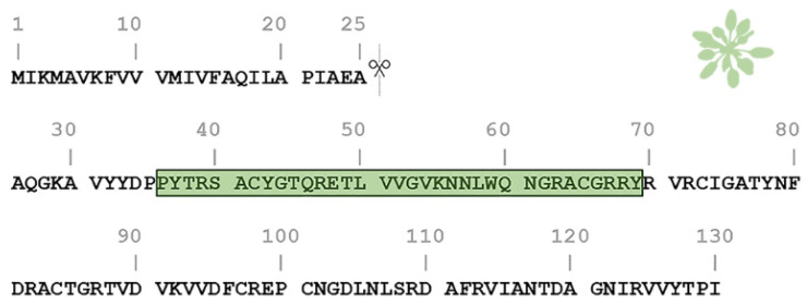
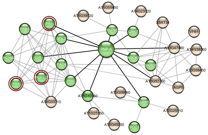
Plant natriuretic peptides (PNPs) are hormones that have been extracted from many different species, with the Arabidopsis thaliana PNP (AtPNP-A) being the most studied among them. AtPNP-A is a signaling molecule that consists of 130 residues and is secreted into the apoplast, under conditions of biotic or abiotic stress. AtPNP-A has distant sequence homology with human ANP, a protein that forms amyloid fibrils in vivo. In this work, we investigated the amyloidogenic properties of a 34-residue-long peptide, located within the AtPNP-A sequence, in three different pH conditions, using transmission electron microscopy, X-ray fiber diffraction, ATR FT-IR spectroscopy, Congo red and Thioflavin T staining assays. We also utilize bioinformatics tools to study its association with known plant amyloidogenic proteins and other A. thaliana proteins. Our results reveal a new case of a pH-dependent amyloid forming peptide in A. thaliana, with a potential functional role.

Keywords: Arabidopsis thaliana; amyloid fibrils; functional amyloid; natriuretic peptides; plant natriuretic peptides.


Conflict of interest statement

The authors declare no conflict of interest.

Figures

Figure 1
Figure 1
Amino acid sequence of the Arabidopsis thaliana plant natriuretic peptide (AtPNP-A). The pre-processed sequence of AtPNP-A (UniProt AC: Q9ZV52) consists of 130 residues and contains a signal peptide in its N-terminal (residues 1–25), responsible for its secretion. The active domain of the AtPNP-A molecule (AtPNP-A36–69) corresponds to the 36–69 region (green box).
Figure 2
Figure 2
Transmission electron micrographs of amyloid-like fibrils derived from AtPNP-A36–69 in three different pH conditions. (A) When incubated at pH 5.6 (MES buffer), the thinnest AtPNP-A36–69 fibrils had a diameter of approximately 70 Å (black arrows). These fibrils interacted, forming either ribbon-like (black arrowheads) or twisted (white arrows) structures. (B) When incubated at pH 7.4 (HEPES buffer), AtPNP-A36–69 formed amyloid fibrils, with a diameter of approximately 70 Å (black arrows). The fibrils formed ribbon-like structures with various widths (black arrowheads). (C) When AtPNP-A is incubated at pH 8.4 (Tris buffer), it formed straight, unbranched and of undefined length fibrils, with a diameter ranging from approximately 90 Å to 100 Å (black arrows). In this case, ribbon-like structures were also observed (black arrowheads). Bar 250 nm.
Figure 3
Figure 3
Structural features of AtPNP-A36–69 fibrils. (A) X-ray diffraction pattern produced by oriented fibers containing AtPNP-A36–69 amyloid fibrils. The pattern is indicative of the “cross-β” structure, displaying both a meridional (M) 4.6 Å and an equatorial (E) 9.8 Å reflection, corresponding to the distance between consecutive β-strands and the distance between packed β-sheets, respectively. The random packing of the fibrils results in the occurrence of rings instead of oriented reflections. (B) ATR FT-IR (1100–1800 cm−1) spectra results, obtained from suspensions of fibrils, produced from the AtPNP-A36–69, cast on a flat stainless-steel plate and left to air-dry slowly, at ambient conditions, to form hydrated, thin films. Second derivative spectra are also included. (see also Table 1).
Figure 4
Figure 4
ThT and Congo Red assays of AtPNP-A36–69 amyloid-like fibrils. (A) ThT fluorescence emission spectrum of AtPNP-A36–69 incubated in three different pH conditions over a period of 10 h. AtPNP-A36–69, when dissolved in pH 7.4 (HEPES buffer) and 8.4 (Tris buffer), reached its peak after approximately 5 h (red and green line, respectively). When AtPNP-A36–69 is incubated in pH 5.6 (MES buffer), reached the highest value after approximately 1 h. Error bars in ThT fluorescence emission spectra represent standard deviation among triplicates. (B) Congo red staining results from the AtPNP-A36–69 fibrils. Photomicrographs of the fibrils (Left) under bright field illumination and (Right) under crossed polars. The typical for amyloid fibrils yellow/green birefringence clearly appears under crossed polars (Right). Bar 300 μm.
Figure 5
Figure 5
The interaction network of AtPNP-A. Interaction data for the creation of this network were gathered from the publicly available database STRING [57]. The network was visualized using Cytoscape 3.7.2 [58], a freely available platform for biological network visualization and analysis. The network consists of 32 nodes and 85 edges. Proteins are depicted as nodes and interactions as edges. Three of the proteins of the network (red circles) are associated with amyloid-forming proteins. Green-colored nodes are proteins that functional enrichment analysis indicated their association with defense responses. Black-colored edges are the interactions of AtPNP-A and grey-colored edges are the interactions of the AtPNP-A first neighbors.

Similar articles

Cited by

References

    1. Dobson C.M. Protein folding and misfolding. Nature. 2003;426:884–890. doi: 10.1038/nature02261. - DOI - PubMed
    1. Chiti F., Dobson C.M. Amyloid formation by globular proteins under native conditions. Nat. Chem. Biol. 2009;5:15–22. doi: 10.1038/nchembio.131. - DOI - PubMed
    1. Cliffe R., Sang J.C., Kundel F., Finley D., Klenerman D., Ye Y. Filamentous Aggregates Are Fragmented by the Proteasome Holoenzyme. Cell Rep. 2019;26:2140–2149.e3. doi: 10.1016/j.celrep.2019.01.096. - DOI - PMC - PubMed
    1. Dobson C.M., Knowles T.P.J., Vendruscolo M. The Amyloid Phenomenon and Its Significance in Biology and Medicine. Cold Spring Harb. Perspect. Biol. 2020;12:a033878. doi: 10.1101/cshperspect.a033878. - DOI - PMC - PubMed
    1. Nastou K.C., Nasi G.I., Tsiolaki P.L., Litou Z.I., Iconomidou V.A. AmyCo: The amyloidoses collection. Amyloid Int. J. Exp. Clin. Investig. 2019;26:112–117. doi: 10.1080/13506129.2019.1603143. - DOI - PubMed