. 2021 Dec 28;11(1):94.
doi: 10.3390/plants11010094.

In Vitro Photoprotective, Anti-Inflammatory, Moisturizing, and Antimelanogenic Effects of a Methanolic Extract of Chrysophyllum lucentifolium Cronquist

Affiliations

In Vitro Photoprotective, Anti-Inflammatory, Moisturizing, and Antimelanogenic Effects of a Methanolic Extract of Chrysophyllum lucentifolium Cronquist

Chaoran Song et al. Plants (Basel). .

Abstract

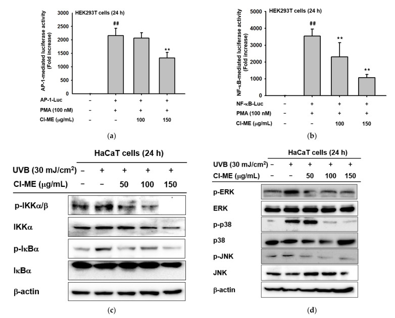
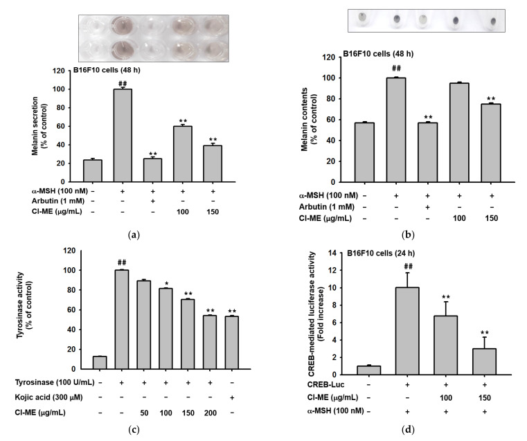
UVB exposure causes DNA mutation and ROS generation, which lead to skin photoaging, skin wrinkling, skin sagging, and uneven skin pigmentation. ROS activate the NF-κB and MAPK signaling pathways leading to production of inflammatory molecules such as COX-2, collagen-degrading proteins such as matrix metalloproteinases (MMPs), and moisture-deficiency-related proteins such as hyaluronidases (HYALs). UVB exposure also induces irregular skin pigmentation though melanin overproduction, related to CREB transcription factor activity and transcription of melanogenesis genes. Here, we demonstrate that Chrysophyllum lucentifolium methanol extract (Cl-ME) has antioxidant activity; it dose-dependently decreased the expression of COX-2, MMP-1, MMP-9, HYAL-1, and HYAL-4 by downregulating the NF-κB (IKKα/β, IκBα) and MAPK (ERK, JNK, and p38) pathways and increased the expression of Col1a1, which encodes a protein important for maintaining skin elasticity. Cl-ME also showed promising antimelanogenic activity by decreasing the expression of CREB, a transcription factor, which in turn inhibited the expression of genes encoding tyrosinase, MITF, TYRP1, and TYRP2. In summary, a methanol extract of C. lucentifolium exhibited antiphotoaging and antimelanogenic activity and could be useful in the cosmeceutical industry.

Keywords: AP-1; NF-κB; ROS; skin aging; skin whitening.


Conflict of interest statement

The authors have no conflict of interest to declare.

Figures

Figure 1
Figure 1
Compound analysis and cytotoxicity of Cl-ME toward HaCaT, HDF, HEK293, and B16F10 cells. (a) Viability of HaCaT, (b) HDF, (c) HEK293T, and (d) B16F10 cells after 24 h treatment with Cl-ME (0–200 µg/mL) was measured using the MTT assay. (e) The phytochemical fingerprinting of Cl-ME was observed by HPLC.
Figure 2
Figure 2
Antioxidant and photoprotective effects of Cl-ME on UVB- and H2O2-induced damage in HaCaT cells. (a,c) Morphological changes in UVB-treated (30 mJ/cm2) HaCaT cells after 24 h treatment with Cl-ME (0–150 µg/mL). (b,d) Survival (% of control) of UVB (b)- or H2O2 (d)-treated HaCaT cells after a 24 h treatment with Cl-ME. (e) Protective activity of Cl-ME against ROS and cell damage as measured through DCFDA (10 μM) and DAPI (1 μL/mL) staining in HaCaT cells after 24 h of treatment. (f) Radical scavenging activity of Cl-ME was measured using the ABTS assay with ascorbic acid as the positive control. ## p < 0.01 compared to the normal group and ** p < 0.01 compared to the control group.
Figure 2
Figure 2
Antioxidant and photoprotective effects of Cl-ME on UVB- and H2O2-induced damage in HaCaT cells. (a,c) Morphological changes in UVB-treated (30 mJ/cm2) HaCaT cells after 24 h treatment with Cl-ME (0–150 µg/mL). (b,d) Survival (% of control) of UVB (b)- or H2O2 (d)-treated HaCaT cells after a 24 h treatment with Cl-ME. (e) Protective activity of Cl-ME against ROS and cell damage as measured through DCFDA (10 μM) and DAPI (1 μL/mL) staining in HaCaT cells after 24 h of treatment. (f) Radical scavenging activity of Cl-ME was measured using the ABTS assay with ascorbic acid as the positive control. ## p < 0.01 compared to the normal group and ** p < 0.01 compared to the control group.
Figure 3
Figure 3
Prevention of moisture loss and collagen degradation and the effect of collagen formation of Cl-ME. (a,b) Expression of COX-2 in UVB (30 mJ/cm2)- or H2O2-(100 µM) treated HaCaT cells after 24 h treatment with Cl-ME. (c,d) Expression of MMP-1, MMP-9, HYAL-1, and HYAL-4 in HaCaT cells after a 24 h treatment with H2O2 and Cl-ME. (e,f) Recovery of Col1a1 expression in UVB-treated (30 mJ/cm2) or H2O2-treated HDF cells after 24 h of treatment with Cl-ME. (f) Recovery of Col1a1 expression in HDF cells after a 24 h treatment with H2O2 and Cl-ME. (g) Expression of the Col1a1 gene in HDF cells after a 24 h treatment with Cl-ME. (h) Col1a1-mediated luciferase activity in HEK293T cells, measured after 24 h of treatment with Cl-ME; retinol was used as the positive control. * p < 0.05 and ** p < 0.01 compared to the control group.
Figure 3
Figure 3
Prevention of moisture loss and collagen degradation and the effect of collagen formation of Cl-ME. (a,b) Expression of COX-2 in UVB (30 mJ/cm2)- or H2O2-(100 µM) treated HaCaT cells after 24 h treatment with Cl-ME. (c,d) Expression of MMP-1, MMP-9, HYAL-1, and HYAL-4 in HaCaT cells after a 24 h treatment with H2O2 and Cl-ME. (e,f) Recovery of Col1a1 expression in UVB-treated (30 mJ/cm2) or H2O2-treated HDF cells after 24 h of treatment with Cl-ME. (f) Recovery of Col1a1 expression in HDF cells after a 24 h treatment with H2O2 and Cl-ME. (g) Expression of the Col1a1 gene in HDF cells after a 24 h treatment with Cl-ME. (h) Col1a1-mediated luciferase activity in HEK293T cells, measured after 24 h of treatment with Cl-ME; retinol was used as the positive control. * p < 0.05 and ** p < 0.01 compared to the control group.
Figure 4
Figure 4
Cl-ME-mediated downregulation of the inflammatory NF-κB and AP-1 pathways. (a) NF-κB-mediated luciferase activity in PMA-activated (100 nM) HEK293T cells, measured after a 24 h treatment with Cl-ME (0–150 µg/mL). (b) AP-1-mediated luciferase activity in PMA-activated (100 nM) HEK293T cells, measured after a 12 h treatment with Cl-ME (0–150 µg/mL). (c) Phosphorylated and total forms of NF-κB pathway proteins (IKKα/β and IκBα) obtained from UVB-treated (30 mJ/cm2) HaCaT cells after a 24 h treatment with Cl-ME (0–150 µg/mL). (d) Phosphorylated and total forms of AP-1 pathway proteins (ERK, JNK, and p-38) from UVB-treated HaCaT cells after a 24 h treatment with Cl-ME (0–150 µg/mL). ## p < 0.01 compared to the normal group and ** p < 0.01 compared to the control group.
Figure 5
Figure 5
Antimelanogenic effect of Cl-ME in murine melanoma B16F10 cells. (a) Melanin secretion in B16F10 cells after 48 h of treatment with Cl-ME (0–150 µg/mL); arbutin (1 mM) was used as the positive control. (b) Melanin content in B16F10 cells after 48 h of treatment with Cl-ME (0–150 µg/mL) with arbutin (1 mM) as the positive control. (c) Tyrosinase activity assay of Cl-ME (0–200 µg/mL) with kojic acid (300 µM) as the positive control. (d) CREB-mediated luciferase activity in α-MSH-activated B16F10 cells after 48 h of treatment with Cl-ME (0–150 µg/mL). (e) Expression of MITF, TRP-1, and TRP-2 proteins in α-MSH-activated B16F10 cells after a 48 h treatment with Cl-ME (0–150 µg/mL). (f) Phosphorylated forms of CREB and total amounts of CREB and MITF in α-MSH-activated B16F10 cells after a 48 h treatment with Cl-ME (0–150 µg/mL); arbutin was the positive control. (g) Phosphorylated forms of tyrosinase obtained from α-MSH-activated B16F10 cells after a 48 h treatment with Cl-ME (0–150 µg/mL) using arbutin as the positive control. ## p < 0.01 compared to the normal group, and * p < 0.05 and ** p < 0.01 compared to the control group.
Figure 5
Figure 5
Antimelanogenic effect of Cl-ME in murine melanoma B16F10 cells. (a) Melanin secretion in B16F10 cells after 48 h of treatment with Cl-ME (0–150 µg/mL); arbutin (1 mM) was used as the positive control. (b) Melanin content in B16F10 cells after 48 h of treatment with Cl-ME (0–150 µg/mL) with arbutin (1 mM) as the positive control. (c) Tyrosinase activity assay of Cl-ME (0–200 µg/mL) with kojic acid (300 µM) as the positive control. (d) CREB-mediated luciferase activity in α-MSH-activated B16F10 cells after 48 h of treatment with Cl-ME (0–150 µg/mL). (e) Expression of MITF, TRP-1, and TRP-2 proteins in α-MSH-activated B16F10 cells after a 48 h treatment with Cl-ME (0–150 µg/mL). (f) Phosphorylated forms of CREB and total amounts of CREB and MITF in α-MSH-activated B16F10 cells after a 48 h treatment with Cl-ME (0–150 µg/mL); arbutin was the positive control. (g) Phosphorylated forms of tyrosinase obtained from α-MSH-activated B16F10 cells after a 48 h treatment with Cl-ME (0–150 µg/mL) using arbutin as the positive control. ## p < 0.01 compared to the normal group, and * p < 0.05 and ** p < 0.01 compared to the control group.
Figure 6
Figure 6
Schematic pathway illustrating the moisturizing, antiaging, anti-inflammatory, and antimelanogenic effects of Cl-ME in vitro. Upon UVB, H2O2, or α-MSH stimulation conditions, AP-1, NF-κB, and CREB pathways are initiated. By these pathways, expression of MMPs, COX-2, HYALs, and tyrosinase is increased, leading to skin inflammation, collagen degradation, and moisturizing deficiency, as well as pigmentation. Cl-ME plays its role in moisturizing, antiaging, anti-inflammatory, and antimelanogenic effects by targeting AP-1, NF-κB, and CREB pathways.

Similar articles

Cited by

References

    1. Kohl E., Steinbauer J., Landthaler M., Szeimies R.M. Skin ageing. J. Eur. Acad. Dermatol. Venereol. 2011;25:873–884. doi: 10.1111/j.1468-3083.2010.03963.x. - DOI - PubMed
    1. Amaro-Ortiz A., Yan B., D’Orazio J.A. Ultraviolet radiation, aging and the skin: Prevention of damage by topical cAMP manipulation. Molecules. 2014;19:6202–6219. doi: 10.3390/molecules19056202. - DOI - PMC - PubMed
    1. Parrado C., Mascaraque M., Gilaberte Y., Juarranz A., Gonzalez S. Fernblock (Polypodium leucotomos extract): Molecular mechanisms and pleiotropic effects in light-related skin conditions. Photoaging Ski. Cancers. 2016;17:1026. - PMC - PubMed
    1. Farhana L., Dawson M.I., Fontana J.A. Apoptosis induction by a novel retinoid-related molecule requires nuclear factor-κB activation. Cancer Res. 2005;65:4909–4917. doi: 10.1158/0008-5472.CAN-04-4124. - DOI - PubMed
    1. Cooper S.J., Bowden G.T. Ultraviolet B regulation of transcription factor families: Roles of nuclear factor-kappa B (NF-kappaB) and activator protein-1 (AP-1) in UVB-induced skin carcinogenesis. Curr. Cancer Drug Targets. 2007;7:325–334. doi: 10.2174/156800907780809714. - DOI - PMC - PubMed